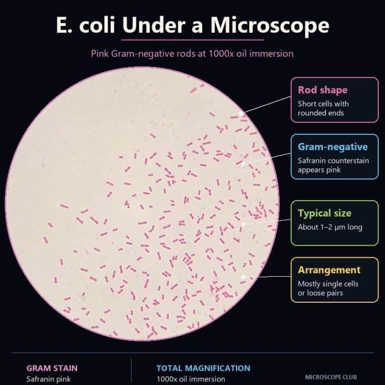
E. coli under a microscope appears as tiny pink, rod-shaped cells, usually scattered singly or in loose pairs against a pale background. You need 1000x total magnification with oil immersion after a Gram stain to actually resolve that shape. A standard light microscope can show you the cell’s shape, size, arrangement, and Gram reaction — but it cannot tell you which strain you’re looking at, or whether it’s dangerous.
How to Prepare and View an E. coli Slide
Getting a clear look at Escherichia coli starts well before your eye reaches the eyepiece. Most of what determines whether you see crisp pink rods or a purple smear happens during slide prep, not focusing.
What You’ll Need
A compound microscope with a 100x oil-immersion objective, a bottle of immersion oil, a basic Gram stain kit (crystal violet, iodine mordant, decolorizer, safranin), and a smear — either a commercially prepared, pre-fixed E. coli slide or a smear made from a non-pathogenic lab strain under proper classroom supervision. If you’re new to slide setup generally, it helps to review how to prepare microscope slides before starting a stain.
Making the Smear and Heat-Fixing
Spread a thin, even film of the culture across the slide — thin enough that you could almost read text through it. A thick smear dries into an opaque purple mass that never resolves into individual rods, so err on the side of too little material rather than too much. Pass the slide (smear-side up) through a flame two or three times to heat-fix it; this kills and anchors the cells so they survive the rinsing steps instead of washing off the glass.
The Gram Stain Steps
- Flood the smear with crystal violet for about 60 seconds, then rinse with water.
- Apply iodine (the mordant) for about 60 seconds, then rinse.
- Decolorize with alcohol for only a few seconds, tilting the slide until the runoff turns clear — stop as soon as it does.
- Counterstain with safranin for 30–60 seconds, then rinse and gently blot dry with bibulous paper.
The decolorizing step is where most beginners lose their result. Over-decolorize and you’ll strip color from everything, including Gram-positive controls; under-decolorize and the whole slide stays purple, masking the true Gram-negative reaction. A few seconds of alcohol, watched closely, is the difference.
Getting It in Focus at 1000x
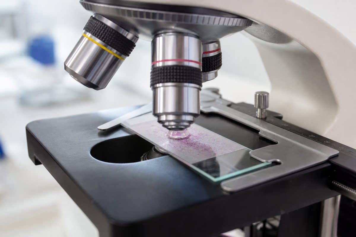
Find the smear at low power first, then switch to the 100x oil-immersion objective — place a single drop of immersion oil directly on the slide and lower the objective into it rather than trying to focus up through air. Total magnification is eyepiece power times objective power, so a 10x eyepiece with the 100x oil objective gives you 1000x, which is the standard needed to actually see E. coli’s rod shape rather than an indistinct speck.
What E. coli Looks Like Under the Microscope
Shape and Arrangement
At 1000x, E. coli reads as tiny pink or magenta rods — like grains of rice shrunk down to specks — often lying in loose clumps or scattered pairs. They’re smaller and plainer than most classroom specimens; if you’ve looked at onion or cheek cells before, expect something noticeably smaller with no visible internal detail. There’s no shading or texture inside an individual cell, just a solid pink rod with rounded ends. That flatness is normal — beginners often assume their scope is malfunctioning when they can’t find internal structures, but a featureless rod is the correct, expected result. E. coli measures roughly 1–2 micrometers long and about 0.5 micrometers wide, which sits close to the resolving power of a light microscope (~0.2 micrometers), so texture and internal features simply aren’t available at this scale.

Color and What the Gram Result Means
E. coli is Gram-negative, which is why it stains pink rather than purple. During the Gram procedure, its thin peptidoglycan wall — sandwiched between an inner membrane and an outer membrane containing lipopolysaccharide — fails to trap the crystal violet-iodine complex once alcohol hits it. The cell loses that purple color and instead picks up the pink safranin counterstain. Gram-positive bacteria, which have a thick peptidoglycan wall and no outer membrane, hold onto the crystal violet and stay purple instead. You can see that contrast directly in a slide of the Gram-positive bacteria in yogurt, which keep their violet color under the same procedure.
What Magnification You Need (400x vs 1000x)
Total magnification equals eyepiece power multiplied by objective power — a 10x eyepiece with a 40x objective gives 400x, and the same eyepiece with a 100x oil objective gives 1000x. E. coli’s size relative to that math is exactly why low power falls short: at 400x you’ll see faint dark or pink specks that confirm something is there, but not the rod shape itself. Only 1000x with true oil immersion resolves individual cells clearly. If you want the underlying math spelled out, see how to calculate total magnification.
| Magnification | What you’ll actually see |
|---|---|
| 100x | Nothing distinguishable — the field looks empty or faintly clouded |
| 400x | Tiny dark or pink specks; you can tell something is present but not its shape |
| 1000x (oil immersion) | Clear pink rods, singly or in pairs, against a pale background |
Handheld or digital microscopes that advertise “1000x” through digital zoom won’t deliver this — without a true 100x oil objective and immersion oil, you’re still limited to the resolution of a much lower-power lens.

What You Can’t See Under a Light Microscope
A Gram-stained slide tells you shape, size, and Gram reaction — nothing more. E. coli is motile via peritrichous flagella (thin, whip-like structures distributed all over the cell surface). Those flagella are far too thin to resolve with an ordinary stain or standard light microscope. Seeing them requires a specialized flagellar stain, which chemically thickens them, or electron microscopy. The same is true of fimbriae (short, hair-like adhesion structures), the texture of the outer membrane, and internal features like the nucleoid. E. coli is a prokaryote with no true membrane-bound nucleus, just a loose region of DNA that a light microscope can’t distinguish from the rest of the cytoplasm. For a look at what those extra layers of detail actually reveal, see what electron microscopes reveal in bacterial cells.
This is also why you can’t identify E. coli by sight alone, and why you can’t tell a harmless strain from a dangerous one under any light microscope. According to the CDC, most E. coli strains are a normal, harmless part of the human gut. A small number — including Shiga toxin-producing strains like O157:H7 — can cause serious foodborne illness (CDC: About E. coli). Both look identical as pink rods on a slide; distinguishing them requires selective culture media (such as MacConkey or EMB agar) and biochemical or molecular testing, not a microscope (NCBI: Gram Staining).
This article is educational, not diagnostic. It explains what a Gram-stained slide shows about E. coli’s appearance — it is not a tool for identifying contamination or infection. Never attempt to culture bacteria from food, water, or other unknown sources at home. School and hobby work should use commercially prepared slides or approved non-pathogenic lab strains under proper supervision, with hands washed and the work area disinfected afterward.
Viewing Live E. coli
Because unstained bacteria are nearly transparent, a standard brightfield scope shows almost nothing without a stain. To see live, unstained E. coli, microscopists use phase contrast microscopy or dark-field microscopy, both of which exaggerate subtle differences in refractive index so transparent cells become visible against the background. A hanging-drop or wet mount preparation is the classic way to observe motility directly. You won’t see individual flagella, but you can watch the cells tumble and swim, which is itself good evidence of flagellar movement. For general technique on setting one up, see how to make a wet mount slide.
A well-focused compound scope with a good oil-immersion objective is essential for any of this work; resources like Nikon’s MicroscopyU are a solid reference if you want to go deeper into objective and immersion-oil technique.
Frequently Asked Questions
Can you tell if E. coli is dangerous by looking at it?
No. Harmless gut strains and pathogenic strains like O157:H7 are visually indistinguishable under any light microscope — telling them apart requires selective culture and lab testing, not appearance.
How big is E. coli in microns?
About 1–2 micrometers long and roughly 0.5 micrometers wide — close to the resolving limit of a standard light microscope, which is why individual cells look like plain, featureless rods.
Can you see E. coli flagella under a microscope?
Not with a standard Gram stain or ordinary light microscope. Flagella are visible only with a specialized flagellar stain that thickens them, or with electron microscopy.
Why does my Gram-stained E. coli look all purple instead of pink?
That usually means the decolorizing step was too short or skipped. Alcohol needs to run clear off the slide before you counterstain, or the crystal violet never gets washed out of the Gram-negative cells.
Can you Gram-stain E. coli on a plastic slide, or do you need glass?
Use glass slides. Plastic doesn’t reliably withstand heat-fixing over a flame and can warp or melt, and many plastics aren’t compatible with the alcohol decolorizer.
How long do prepared E. coli slides last before the stain fades?
A properly sealed, commercially prepared slide can last for years if stored away from direct light and heat, though the safranin pink can fade or look uneven over time — that’s normal aging of the stain, not a technique problem.
Conclusion
What E. coli looks like under a microscope comes down to a short, honest list. Tiny pink rods, roughly 1–2 micrometers long, visible in real detail only at 1000x oil immersion after a proper Gram stain. That’s genuinely useful — it confirms shape, size, and Gram reaction — but it’s also the ceiling of what light microscopy can tell you. Flagella, fine surface structures, and strain identity all sit beyond that ceiling, waiting for a flagellar stain, electron microscopy, or a culture plate.
Have you run a Gram stain on E. coli yourself, in a classroom or at home with a prepared slide? Tell us what your decolorizing step looked like, or ask a question in the comments if your slide isn’t turning out the way you expected.
Originally posted 2026-07-27 12:00:00.