Total microscope magnification is one of the first things you need to work out in any biology lab — and it comes down to a single multiplication. Multiply the power of your objective lens by the power of your eyepiece lens and you have your answer. Every compound microscope works the same way, whether you are using a student scope at 40x or pushing a research instrument to 1000x.
The Core Formula and Quick-Reference Table
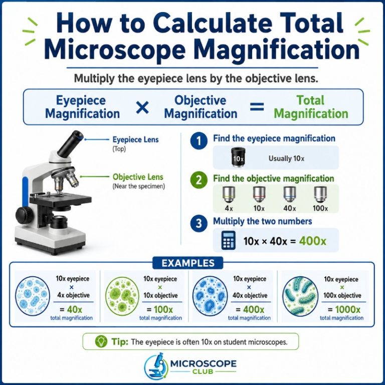
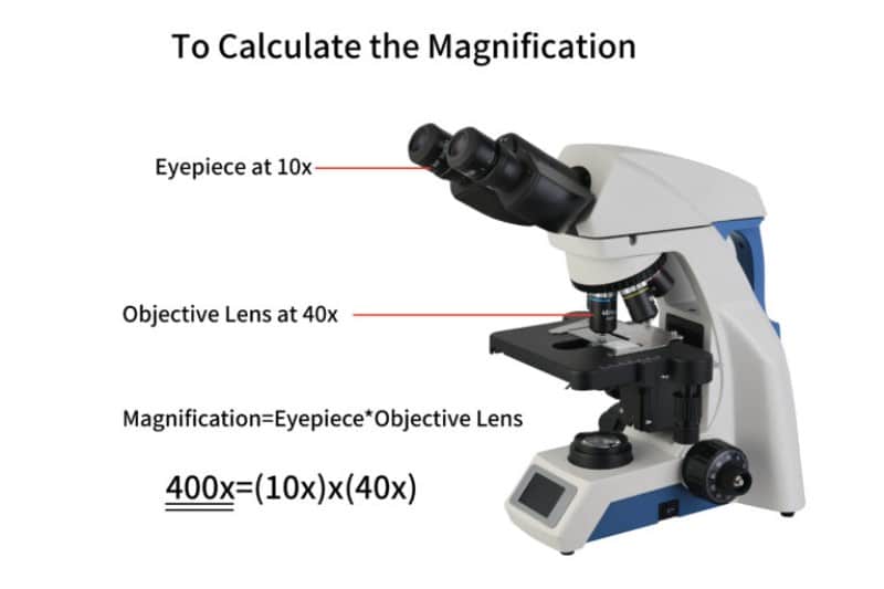
Total microscope magnification is calculated by multiplying the power of the objective lens by the power of the eyepiece lens: Total Magnification = Objective Lens Power × Eyepiece Power. On a standard compound microscope with a 10x eyepiece, switching from the 4x scanning objective to the 40x high-power objective takes you from 40x total magnification all the way to 400x — a tenfold jump by simply rotating the nosepiece. Every microscope works the same way: identify the two numbers engraved on your lenses, multiply them, and you have your answer.
| Objective Lens | × 10x Eyepiece | × 15x Eyepiece |
|---|---|---|
| 4x (scanning) | 40x | 60x |
| 10x (low power) | 100x | 150x |
| 40x (high power) | 400x | 600x |
| 100x (oil immersion) | 1000x | 1500x |
The Total Magnification Formula Explained
A compound light microscope achieves high magnification through two separate lens systems working in series. Understanding what each lens does makes the math intuitive — and prevents the most common mistakes students make in the lab.
What the Eyepiece Lens Does
The eyepiece — also called the ocular lens — is the lens you press your eye against at the top of the microscope. Its job is to act as a magnifying glass for the intermediate image already formed by the objective lens below. Most student and laboratory microscopes come fitted with a 10x eyepiece, which is why 40x, 100x, 400x, and 1000x are the four magnification levels you see quoted most often. Some microscopes ship with a 15x eyepiece, which boosts each tier significantly; a small number of specialty scopes use 5x or 20x oculars. The magnification value is engraved on the top of the eyepiece barrel — look for a marking like “WF10×” or simply “10×.”
What the Objective Lens Does
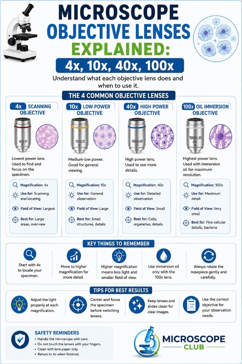
The objective lens is the small lens positioned closest to the specimen, mounted on the rotating nosepiece. It is the primary magnifier in the optical chain — responsible for capturing light from the specimen and producing that first, magnified intermediate image. The parts of a compound microscope include four standard objectives:
- 4x — scanning objective: Lowest power; gives the widest field of view. Use it first to locate your specimen on a slide.
- 10x — low-power objective: Good for examining larger structures and getting oriented at the cellular level.
- 40x — high-power objective: The workhorse for most biology lab work — detailed cell structures become visible.
- 100x — oil-immersion objective: Requires a drop of immersion oil between the lens tip and the coverslip to maintain resolution. Used for bacteria, blood cells, and fine subcellular detail.
The objective’s power is engraved on its barrel. You will often see a second number as well — for example, “40×/0.65.” That second figure (0.65) is the numerical aperture (NA), which describes the lens’s light-gathering ability and is a key factor in microscope resolution and numerical aperture. Do not multiply it into your magnification calculation. Numerical aperture is not a magnification value.
Why You Multiply, Not Add
The reason the formula is multiplication rather than addition comes down to how sequential magnification works. The objective lens enlarges the specimen’s image, say, 40 times its actual size. The eyepiece then takes that already-enlarged image and magnifies it a further 10 times. The total enlargement is 40 × 10 = 400x — not 40 + 10 = 50x. Each lens stage acts on the output of the previous stage, so the effects compound multiplicatively.

Step-by-Step: Calculating Your Microscope’s Magnification
Follow these three steps with any compound microscope:
- Read the eyepiece power. Look at the top of the eyepiece barrel. Find the magnification marking — most commonly “10×.” Write that number down.
- Read the active objective power. Look at the objective currently rotated into position above the stage. Find its engraved magnification (4×, 10×, 40×, or 100×). Ignore any other numbers such as numerical aperture.
- Multiply the two values. Eyepiece power × objective power = total magnification. That’s it.
Example: eyepiece reads 10×, active objective reads 40× → 10 × 40 = 400x total magnification.

Standard Magnification Combinations: Quick-Reference Table
The table below covers every common objective at both 10x and 15x eyepieces. Most competitors only publish the 10x column — having both lets you use the same reference regardless of which eyepiece your scope came with.
| Objective | Name | 10x Eyepiece | 15x Eyepiece | Typical Use |
|---|---|---|---|---|
| 4x | Scanning | 40x | 60x | Locating specimen; overview of tissue sections |
| 10x | Low power | 100x | 150x | General cell structure; plant cross-sections |
| 40x | High power | 400x | 600x | Individual cells; organelles; bacteria colonies |
| 100x | Oil immersion | 1000x | 1500x | Bacteria; blood smears; flagella; fine cell detail |
Note that 1500x sits at or beyond the practical resolution ceiling for visible-light microscopy (more on this below). The 15x + 100x combination is technically possible but yields empty magnification — a bigger image that is no sharper.
Worked Examples
Three common scenarios you will encounter in a biology lab or at home:
Example 1: 4x objective + 10x eyepiece
10 × 4 = 40x. At this low magnification, you are looking at the big picture — entire tissue cross-sections, the layout of leaf veins, or the surface pattern of a fabric fiber. Field of view changes with magnification: at 40x you see a wide area; at 1000x you see a tiny patch of the same slide.
Example 2: 40x objective + 10x eyepiece
10 × 40 = 400x. This is the most-used setting in high school and college biology labs. Individual cells are clearly visible, nuclei are distinguishable, and you can see organelle outlines in larger cells like onion or cheek cells. Preparing a slide for viewing properly matters most at this level — debris or air bubbles are very obvious.
Example 3: 100x objective + 15x eyepiece
15 × 100 = 1500x. This pushes a light microscope to — and slightly past — its practical resolution limit. You will need immersion oil on the 100x objective. The image may look magnified but lack crispness compared to 1000x, because the diffraction limit of visible light (~200 nanometers) cannot be overcome by adding more magnification.

Compound vs. Stereo (Dissecting) Microscope Magnification
Everything above applies to compound microscopes, which are designed for viewing thin, transparent specimens on glass slides. A dissecting (stereo) microscope works on the same optical principle — total magnification = objective × eyepiece — but the numbers look very different in practice.
Stereo microscopes typically deliver 10x–40x total magnification, and sometimes up to 45x or 90x on zoom models. That is far lower than a compound scope’s 40x–1000x range, for a good reason: stereo microscopes are built for 3D surface viewing of coins, insects, circuit boards, and biological specimens that cannot be sliced thin. They use two separate optical paths (one for each eye) to create a stereoscopic image, which requires larger working distances and less extreme magnification. Some stereo scopes have a zoom knob, and in that case total magnification = zoom value × eyepiece × any auxiliary (Barlow) lens attached to the base.
If you are comparing types of microscopes, the key takeaway is: for cellular detail, use a compound scope at high magnification; for intact 3D specimens, use a stereo microscope at low magnification.
Magnification Is Not Resolution — Avoid This Mistake
This is the most important concept that calculator-only guides leave out. Magnification tells you how much larger the image appears relative to the real object. Resolution tells you the smallest gap between two points that the microscope can still show as two distinct points rather than a blurred smear. Resolution is governed by the numerical aperture of the objective lens and the wavelength of light used — not by magnification.
Visible light has a wavelength of roughly 400–700 nanometers. Ernst Abbe’s diffraction limit sets the theoretical resolution of a light microscope at approximately 200 nanometers (0.2 micrometers). In practical terms, a standard compound microscope reaches its useful ceiling at around 1000x total magnification. High-NA oil-immersion setups can push to 1500x–2000x before detail stops improving, but beyond that ceiling you get empty magnification: the image grows larger, but no new detail emerges — blurry structures just become larger blurry structures. You can read more about this at Britannica’s overview of the microscope.
This is why electron microscopes exist. Scanning and transmission electron microscopes use electrons instead of visible light. This yields wavelengths thousands of times shorter and resolutions down to the sub-nanometer range — well beyond what any light microscope can achieve. You can read more about the highest magnification a microscope can achieve to understand where different microscope technologies sit on the resolution spectrum.
The practical implication for lab work: if your 400x image looks sharp and your 1000x image looks fuzzy and washed out, you have not made a mistake — you have hit the physics ceiling. Trying to push to 2000x with a 20x eyepiece will not help.
Common Mistakes When Calculating Magnification
- Forgetting the eyepiece multiplier. The single most common error. Reporting “40x magnification” when you have a 40x objective and a 10x eyepiece means you are off by a factor of 10. The correct answer is 400x.
- Confusing numerical aperture with magnification. The “0.65” engraved after “40×/0.65” on an objective is the numerical aperture — a measure of light-gathering, not power. Do not include it in your magnification calculation.
- Assuming higher magnification always gives a better image. As covered above, magnification beyond ~1000x on a light microscope yields empty magnification. Higher power also narrows the field of view and reduces depth of focus, making it harder to find and track moving specimens.
- Multiplying in a third lens that is not there. Standard compound microscopes have exactly two lens systems in the magnification chain: eyepiece and objective. Only scopes with a Barlow lens, auxiliary lens, or a non-standard tube-lens factor need a third number multiplied in. Check your microscope’s manual if you are unsure.
- Using the wrong objective. Always confirm which objective is locked into position before recording magnification. The 40x and 4x objectives can look similar when you glance quickly at the nosepiece.
Frequently Asked Questions
Why is my microscope blurry at high magnification?
There are a few common reasons: (1) Empty magnification — you have exceeded the ~1000x resolution ceiling of visible light. (2) Missing immersion oil — the 100x oil-immersion objective requires a drop of immersion oil; without it, image quality degrades severely. (3) Focus drift — at high power, even a tiny vibration or touch to the stage throws the image out of focus. Use the fine-focus knob in small increments. (4) Dirty lenses — fingerprints or residue on the eyepiece or objective are magnified along with your specimen.
How do I clean my microscope objective lenses without scratching them?
First use a blower (or gentle puff of breath) to clear loose dust, since dragging grit across glass scratches the coating. Then wipe with lens paper lightly moistened — not soaked — in distilled water or lens-cleaning solution, working in a spiral from the centre outward so dirt is pushed to the edge. Never wipe with dry tissue or use a zig-zag motion, and avoid cotton swabs on coated optics where the stick can scratch the surface.
What is immersion oil, is it safe, and how do I clean it off afterward?
Immersion oil is a clear, high-refractive-index oil placed between the coverslip and the 100x oil-immersion objective so light isn’t lost at the glass-air gap — it should only ever be used on objectives marked “oil.” It is low-hazard but should be wiped off immediately after use, because dried oil is hard to remove and can creep into the microscope’s internal mechanics over time. Clean it with lens paper dampened in a lens-safe solvent (such as anhydrous or blended alcohol), wiping centre-to-edge and using a fresh section of paper on each pass until no residue remains.
How do I prepare a wet-mount slide correctly?
Place a small drop of water (or saline) in the centre of a clean slide — slightly smaller than your coverslip — and set the specimen into the drop so it is fully immersed. To avoid trapping air bubbles, hold the coverslip at about a 45-degree angle with one edge touching the slide beside the drop, then lower it slowly so the liquid spreads and pushes air out ahead of it. Blot any liquid that seeps past the coverslip edge with the corner of a paper towel.
Can I get more magnification just by swapping the eyepiece, or are there other options?
Yes, fitting a higher-power eyepiece (e.g. 15x instead of 10x) raises total magnification, but only up to the microscope’s useful resolution limit — past roughly 1000x on a light microscope you gain only empty magnification. You can also add a Barlow (auxiliary) lens, typically sold in 0.5x to 2.0x factors, which sits in the light path and multiplies the total: a 2x Barlow turns a 100x setup into 200x. Note that lower-than-1x Barlow lenses are also useful because they increase working distance and depth of field rather than power.
Conclusion
Calculating total microscope magnification comes down to one multiplication: objective power × eyepiece power. With a 10x eyepiece — the standard on almost every student scope — you get four working magnification levels: 40x, 100x, 400x, and 1000x. Understanding that each lens stage multiplies the previous one (not adds to it), knowing where to read the numbers on each lens barrel, and recognizing the hard ceiling that resolution places on useful magnification will take you from guessing to confident use of any compound or stereo microscope. A simple microscope (single lens) is the only exception — there, total magnification equals the single lens power alone. For a deeper dive into how microscope optics work across different instrument types, Khan Academy’s microscopy unit is a solid free resource.
Have you tried calculating the magnification on your own microscope? Did the numbers match what you expected — or did you discover your scope had a 15x eyepiece instead of a 10x? Share your setup or any questions in the comments below. We read every one.