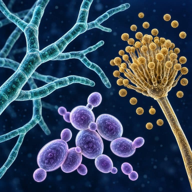
Fungi are a separate kingdom of life, distinct from plants, animals, and bacteria. Under a compound microscope they reveal three signature body forms: thread-like hyphae, budding yeast cells, and spore-bearing structures. Most of that detail comes into focus between 100x and 400x total magnification, especially once a sample is stained. This guide covers what actually makes something a fungus, how to prepare a slide yourself, and what each body form looks like once it’s in view.
What Makes a Fungus a Fungus?
A fungus is defined by four traits working together: a chitin cell wall, a eukaryotic cell structure, absorptive feeding, and a body built from thread-like hyphae. Fungi belong to their own kingdom inside the domain Eukarya, meaning every fungal cell has a membrane-bound nucleus and organelles, just like a plant or animal cell. This is what separates fungi from bacteria, which are prokaryotes with no nucleus at all. The single most useful identifying fact is the cell wall: it’s built from chitin, the same tough polymer found in insect exoskeletons, not the cellulose that stiffens plant cell walls. According to the UC Museum of Paleontology’s introduction to the fungi, this chitin wall combined with a total lack of chlorophyll is what keeps fungi out of the plant kingdom. That’s despite centuries of being lumped in with them. OpenStax summarizes the characteristics that define fungi, including chitin walls and absorptive feeding.
Fungi vs Plants, Animals & Bacteria
Side by side, the four kingdoms split apart cleanly on just a handful of traits. Those traits are cell wall material, whether the cell has a nucleus, how it gets its food, and whether it photosynthesizes.
| Trait | Fungi | Plants | Animals | Bacteria |
|---|---|---|---|---|
| Cell wall | Chitin | Cellulose | None | Peptidoglycan |
| Nucleus (eukaryote?) | Yes | Yes | Yes | No |
| Nutrition mode | Absorptive heterotroph | Autotroph | Ingestive heterotroph | Varies |
| Photosynthesis | No | Yes | No | Rare |
| Example | Mold, yeast, mushroom | Fern, oak tree | Amoeba, insect | E. coli |
How Fungi Feed and Reproduce
Fungi feed by absorption rather than ingestion: they secrete digestive enzymes onto whatever they’re growing on, then absorb the dissolved nutrients back through the cell wall. Most fungi are saprotrophs that break down dead organic matter. Some form parasitic relationships instead, and others, like the fungi in a lichen or a mycorrhiza, live in partnership with an algae or a plant’s roots. Reproduction happens mainly through spores, produced either sexually or asexually on dedicated structures such as sporangia (spore sacs) or conidiophores (stalks bearing chains of spores). The Britannica entry on fungi is a useful reference here — it notes that a single mold colony can release millions of these spores. That’s exactly why mold appears seemingly overnight on stored food.
How to Prepare and View Fungi Under a Microscope
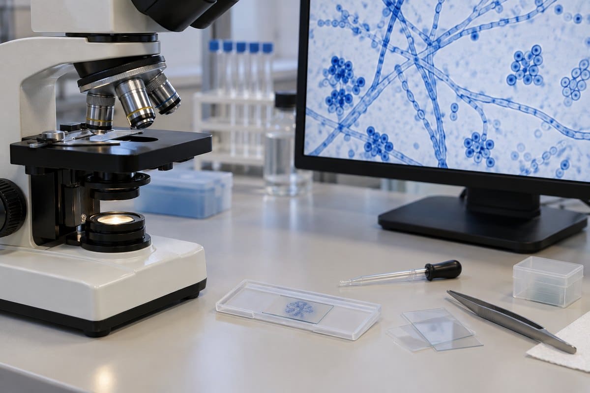
A basic wet mount slide is all you need to see fungal structure clearly, and the whole process takes under five minutes once you’ve got the sample in hand.
What You’ll Need
- A compound microscope with at least a 40x and a 400x objective
- Glass microscope slides and coverslips
- A stain — lactophenol cotton blue or methylene blue both work well
- A sample source: a pinch of visible mold fuzz, a scraping from bread mold, or a pinch of dry active yeast rehydrated in water
- Fine forceps or a piece of clear tape for lifting delicate structures
Step-by-Step Wet Mount
- Collect a small sample. For mold, press a strip of tape lightly onto the fuzzy colony rather than scraping it — this keeps the spore-bearing stalks intact instead of shredding them.
- Place the sample flat in the center of a clean slide.
- Add a drop of stain directly onto the sample. Cotton blue or methylene blue both add contrast to otherwise near-transparent structures.
- Lower the coverslip at an angle, edge first, and let it fall gently onto the drop — pressing straight down flattens and scatters delicate spore heads.
- Focus on low power (40x–100x) first to locate a good area, then switch to 400x to bring individual hyphae or spores into sharp detail.
Best Magnification and Settings
Total magnification between 100x and 400x is where fungal hyphae and spores are best resolved — 1000x oil immersion is rarely necessary just to identify structure. Lighting matters as much as magnification: fungal tissue is naturally translucent, so a fully open diaphragm washes the detail out completely. Stopping down the condenser, or lowering the light intensity, adds the contrast that stain alone doesn’t always provide. If you want a full walkthrough of the mounting technique itself before you start, the site’s guide to how to make a wet mount slide covers the general method. This section builds on that.
What Fungi Look Like Under a Microscope
Once a slide is in focus, fungi show up as one of three recognizable forms — threads, single ovals, or spore clusters — depending on which structure you’ve sampled. Its classification guide provides additional examples of fungal hyphae and spore-producing structures.

Hyphae and Mycelium
Hyphae are the fine, near-transparent threads that make up a fungus’s main body. Under the microscope they look like tangled fishing line or thin cracked-glass filaments running in every direction. A tangled mat of them is called a mycelium. Once cotton blue stain is added, they turn a clean sky-blue against the pale background, and the internal structure becomes obvious. Septate hyphae show faint dark cross-lines dividing them like the joints in a bamboo stalk. Aseptate (coenocytic) hyphae, as in bread mold, look like one smooth continuous pipe with no divisions at all. The most common beginner mistake here is leaving the diaphragm wide open — the light washes out these transparent threads so completely that new observers assume the slide is empty. Stopping down the light, or adding stain, makes them appear almost instantly.
Yeast Cells
Yeast cells appear as small translucent ovals, usually 5–10 micrometers across in a species like Saccharomyces cerevisiae, often clustered together rather than scattered singly. The clearest giveaway that you’re looking at yeast rather than another cell type is a small bump on one side — a daughter cell budding off. Sometimes it looks like a tiny snowman head attached to the parent cell. A few cells may still be joined in short chains where budding hasn’t fully separated them yet.
Spores and Spore-Bearing Structures
Spores show up as tiny, uniform dark specks that visibly drift if the slide is nudged, and they’re produced on distinct stalked structures that are worth learning to tell apart. In Aspergillus, spores radiate outward from a swollen tip in a pattern that looks like a tiny firework or sunburst. That distinctive silhouette is covered in more detail in the site’s guide to identifying Aspergillus under a microscope. In Penicillium, the stalk branches instead into a brush-like cluster, which is exactly what’s described in the article on the brush-shaped conidiophore of Penicillium. The mistake that ruins most first attempts at this view is handling the sample too roughly. Scraping instead of a gentle tape lift tears the conidiophore apart, leaving a pile of loose spores with no intact structure left to identify.

A quick safety note: this is educational content, not medical advice. Most molds are harmless to view briefly on a slide, but some, including certain Aspergillus species, can cause respiratory illness in people who inhale large quantities of spores. Work with small, fresh samples, avoid deliberately disturbing large mold colonies, wash your hands afterward, and don’t attempt to culture an unknown mold in bulk at home. The CDC’s overview of aspergillosis is a good reference if you want to understand the actual risk level involved.
Common Fungi to Observe (and Where to See Them on This Site)
Once you can recognize hyphae, yeast, and spores in general, the fungi below are the easiest starting specimens. Each has its own dedicated guide on this site with sourcing tips and detailed photos.
- Mold — the most accessible specimen of all, often growing on forgotten bread or fruit. See what mold looks like under a microscope for sourcing and slide tips.
- Bread mold — usually Rhizopus, notable for its aseptate hyphae and large black sporangia. Covered in the guide to bread mold and its spore sacs under a microscope.
- Yeast — the easiest single-celled fungus to prepare, since a packet of dry active yeast rehydrated in warm water is ready to mount in minutes. See the guide to observing yeast cells under a microscope.
- Aspergillus — recognizable by its sunburst-shaped conidiophore; full identification detail in the Aspergillus identification guide.
- Penicillium — the brush-shaped counterpart to Aspergillus; see the dedicated Penicillium guide for how to tell the two apart at a glance.
Frequently Asked Questions
Why can’t I see anything on my fungal slide at first?
Fungal tissue is nearly transparent, and a wide-open diaphragm washes out the little contrast it has. Close down the diaphragm or lower the light intensity before assuming the slide is empty — that single adjustment is usually what reveals hyphae or spores that were there all along.
Do you have to stain a sample to identify fungi?
No, but it helps considerably. Hyphae, yeast, and spores are visible unstained under the right lighting, but a stain like lactophenol cotton blue or methylene blue adds contrast that makes structures like septa and spore walls far easier to distinguish, especially for a beginner.
What stain is used for fungi under the microscope?
Lactophenol cotton blue is the standard fungal stain — it colors chitin a clear blue and helps preserve the sample. Methylene blue or iodine also work fine for a quick classroom mount.
Is mold a fungus?
Yes. Mold is simply a common name for certain fast-growing, fuzzy-looking fungi — it isn’t a separate biological category. See the mold under a microscope guide for a closer look.
Is it safe to look at mold under a microscope at home?
Generally yes, if you work with a small, fresh sample on a sealed slide and wash your hands afterward. Avoid disturbing large mold colonies directly or inhaling spores, since some species can affect people with allergies or weakened immune systems.
What’s the difference between mold and mildew?
They’re both common names for fungi rather than distinct scientific groups. “Mildew” is typically used for a thin, surface-level fungal growth (like on damp fabric or leaves), while “mold” usually describes a thicker, fuzzier colony. Under the microscope, both show the same hyphae and spore structures described above.
Conclusion
Fungi earn their own kingdom for good reason: a chitin cell wall, absorptive feeding, and a body built from hyphae set them apart from plants, animals, and bacteria at every level. That holds right down to what you see under the lens. Once you know to look for threads, budding ovals, or radiating spore heads, a slide that looked like an empty smear at low power suddenly makes sense. It resolves into one of those three unmistakable shapes.
Have you tried mounting a mold or yeast sample yourself? Tell us what body form you spotted first — and if you got a clean shot of a spore-bearing structure, share what species you think it was in the comments below.