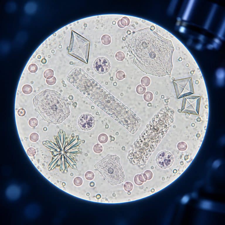
Urine under a microscope reveals a hidden landscape of cells, crystals, and casts that a chemical dipstick test alone can never show. In a microscopic urinalysis, a laboratory technician spins a urine sample down to its sediment and scans that residue under magnification, cataloging exactly which formed elements are present and in what number. This article explains what a technician looks for and why — it’s educational information about the process, not a tool for interpreting your own results, which only a clinician can do.
Here’s what shows up in urine sediment, at a glance:
| Element | What It Looks Like | Why the Lab Notes It |
|---|---|---|
| Red blood cells | Smooth, colorless discs, about 7 µm, no nucleus | Presence flags hematuria — a finding, not a diagnosis |
| White blood cells | Grainy, about 12 µm, with a lobed nucleus | Higher counts flag pyuria, often tied to inflammation |
| Epithelial cells | Large flat squamous cells, rounder transitional cells, tiny renal tubular cells | Type and number point to collection technique or tissue origin |
| Casts | Cylindrical molds — clear, granular, cellular, or waxy | Reflect conditions inside the kidney tubule where they formed |
| Crystals | Envelope, coffin-lid, or rhombic shapes, depending on pH | Shape reveals urine pH and composition; most are harmless |
| Microorganisms | Rods, cocci, budding yeast, or motile organisms | May signal infection or a contaminated collection |

From Sample to Slide — How Urine Is Prepared for Microscopy
A microscopic urinalysis starts with a fresh, clean-catch midstream sample, examined within about two hours or refrigerated until it can be. That timing matters more than it sounds: cells lyse, casts dissolve, and new crystals crash out of solution the longer urine sits. A stale sample shows the lab artifacts of storage rather than what was actually in the body. Refrigeration alone is enough to precipitate a fine cloud of amorphous phosphates or urates — a dusty field that looks alarming but is really just cold, not disease. The NCBI Bookshelf describes preparing and examining urinary sediment under low and high power.
The technician centrifuges roughly 10 to 15 mL of urine at low speed — around 400 g, or 1,500 to 2,000 rpm — for about five minutes. The supernatant gets decanted off, and the button of sediment left at the bottom of the tube is resuspended and placed as a single drop under a coverslip. That button settles back down fast once it’s mixed, so the mount has to happen quickly before elements start clumping again. This is a lab procedure, not a home test — it’s described here to explain what shows up under the lens, not as an instruction set.
Because most of what’s in urine sediment is unstained and low-contrast, the technician closes down the condenser diaphragm to cut the light rather than flooding the field. Too much light washes out hyaline casts and red cells entirely, so the field should look almost dim before the pale, low-refractive elements come into view. A drop of Sternheimer-Malbin stain or a switch to phase-contrast optics can sharpen the same low-contrast structures further.
Low Power vs. High Power: What Each Pass Is For
The exam happens in two passes. The technician scans first at low power field — 100× total magnification, using a 10× objective. This first pass sweeps the edges of the coverslip rather than the center, since casts tend to drift outward and settle at the border. Casts get counted per low power field. The second pass moves to high power field, 400× total using a 40× objective, for the finer detail: cells, crystals, and bacteria, which are counted per high power field. Skipping straight to high power is a common beginner mistake, because it misses the casts entirely. They’re large enough to spot at low power but easy to overlook once the field of view narrows.
The Cells — Red Blood Cells, White Blood Cells, and Epithelial Cells
Red blood cells are the plainest objects in the field: smooth, colorless, biconcave discs with no nucleus. They measure roughly the same 7 µm across as red blood cells seen under the microscope anywhere else in the body. In concentrated urine they crenate into spiky little balls; in dilute or alkaline urine they swell and lyse into faint “ghost” rings that are almost transparent. A normal count runs about 0 to 2 per high power field. This is exactly what “microscopic hematuria” means — blood cells visible only under the lens, with the urine looking perfectly normal to the naked eye. MedlinePlus explains how microscopic hematuria can be detected during urinalysis.

White blood cells, mostly neutrophils, are larger and grainier — about 12 µm, with a granular cytoplasm and a lobed nucleus. That lobed nucleus makes them easy to tell apart from red cells at a glance, the same way how the five white blood cell types are told apart in a stained blood smear. Normal is roughly 0 to 5 per high power field; an elevated count is called pyuria. A frequent point of confusion for beginners is mistaking budding yeast for red or white cells. Yeast is oval, shows a visible bud, and doesn’t lyse the way a red cell does.
Epithelial cells come in three distinct shapes. Squamous cells are the largest and most common — flat, angular sheets that look almost like crumpled cellophane or a fried egg, with a tiny speck of a nucleus. They shed from the urethra or genital tract. In high numbers they usually flag a collection issue rather than disease. Transitional (urothelial) cells are smaller and round-to-pear-shaped, coming from the bladder or ureters. Renal tubular cells are the smallest of the three, round, with a proportionally large nucleus, and originate from the kidney tubule itself. They’re similar in scale to the flat squamous epithelial cells like these cheek cells most students examine first in a biology lab.
Casts — The Kidney’s Fingerprints
A urine cast is a cylindrical mold of the kidney tubule that formed it, built on a matrix of Tamm-Horsfall protein, also called uromodulin. Because a cast takes the exact shape of the tubule lumen, its presence at all tells the lab the material came from inside the kidney rather than lower down the urinary tract. Whatever gets trapped in that protein matrix as it hardens tells the lab which type of cast it is.

Hyaline casts are clear, colorless, and have such a low refractive index that they’re nearly invisible. Dimming the light is what makes them pop, and they’re found by sweeping the coverslip’s edges rather than staring at the center. A few hyaline casts can be a normal finding after exercise or mild dehydration. Granular casts show a fine-to-coarse grainy texture inside the same cylindrical shape. Cellular casts contain intact red cells, white cells, or epithelial cells, and the identity of the trapped cell points the lab toward where in the kidney the cast originated. Waxy casts have sharp, well-defined edges and a high refractive index, while fatty casts contain visible lipid droplets.
Crystals — Shapes That Depend on pH
Crystal shape in urine sediment is a direct function of pH, not of any single disease. In acidic urine, calcium oxalate crystallizes in two forms. The dihydrate form looks like a small sealed envelope — a square with an X across it that catches the light and sparkles under the objective — while the monohydrate form is more of a dumbbell or oval. Uric acid crystals in acidic urine appear as yellow-brown rhombic plates or rosette clusters.
In alkaline urine, triple phosphate (struvite) crystals form distinctive three-dimensional “coffin lid” prisms with beveled ends. Amorphous phosphates show up as a fine granular dust, and ammonium biurate forms spiky “thorn-apple” spheres. Most crystals seen this way are simply diet- or pH-related and benign. A smaller set — cystine hexagons, notched cholesterol plates, tyrosine, leucine, and certain drug crystals — are clinically notable rather than routine. Calcium oxalate and uric acid crystals in particular are the same mineral types documented in what kidney stone crystals look like under a microscope, since kidney stones and urine crystals share a chemical origin. According to the National Institute of Diabetes and Digestive and Kidney Diseases, urinalysis findings like these are one of the ways clinicians evaluate the mineral composition behind kidney stones. Identifying a crystal type on a slide, though, is a lab observation, not a stone diagnosis on its own.
Bacteria, Yeast, and Other Microscopic Finds
Bacteria in urine sediment show up as rods or cocci, sometimes visibly motile at 400×. At first glance they can look like shimmering dots or debris, until the ones that twitch give themselves away. According to the StatPearls clinical reference on urinalysis, E. coli is the organism most often identified this way. It’s the same bacterium covered in more depth in E. coli, the most common urinary organism, under the microscope. Yeast, usually Candida, appears as oval cells showing budding and sometimes forming thread-like pseudohyphae; Trichomonas vaginalis is identifiable by its motile, pear-shaped body and whip-like flagella.
The sediment can also hold mucus threads, occasional spermatozoa, and artifacts that have nothing to do with the patient at all — cotton or clothing fibers, starch or talc granules, and oil droplets. Air bubbles trapped at a drying coverslip edge are a common one too, which beginners sometimes mistake for cells because they form perfect dark rings.
What a “Normal” Microscopic Exam Looks Like
Reference ranges give a sense of scale, though every lab sets its own and only a clinician interprets what a given result means for an individual patient. General ranges like these are published by resources such as MedlinePlus (NIH):
| Element | Typical Reference Range |
|---|---|
| Red blood cells | 0–2 per high power field |
| White blood cells | 0–5 per high power field |
| Casts | 0 to a few hyaline casts per low power field |
| Epithelial cells | Few squamous cells; renal tubular cells rare |
| Crystals | None to few, usually pH-related and benign |
These counts and the elements behind them are reported in standardized units — per high power field for cells, per low power field for casts. Raw numbers only mean something in that specific context. A result outside a typical range is a data point for a clinician to evaluate, not a self-contained answer.
Frequently Asked Questions
Can artifacts like fibers or air bubbles be mistaken for real urine sediment elements?
Yes — cotton or clothing fibers, starch or talc granules, and air bubbles trapped at a drying coverslip edge are common contaminants. Air bubbles in particular form perfect dark rings that beginners sometimes mistake for cells, which is why a technician cross-checks size, shape, and texture against known reference elements before recording a finding.
Is blood in urine visible under a microscope even if you can’t see it by eye?
Yes. Microscopic hematuria specifically means red blood cells are detected under magnification while the urine looks completely normal in color to the naked eye. Visible, eye-detectable blood in urine is a separate and more obvious finding.
Can you do a urinalysis at home with a microscope?
A hobbyist microscope can show you cells and crystals in a urine sample for educational observation, but it cannot replace a clinical urinalysis. Accurate identification depends on controlled centrifugation, calibrated optics, and trained interpretation — and any real health question belongs with a clinician, not a home setup.
What’s the difference between a granular cast and a cellular cast?
Both are cylindrical molds built on the same Tamm-Horsfall protein matrix, but a granular cast shows a fine-to-coarse grainy texture with no identifiable cell structure inside. A cellular cast, by contrast, contains intact red cells, white cells, or epithelial cells that the lab can identify by type — pointing to a more specific origin inside the kidney.
How is microscopic urinalysis different from a dipstick urine test?
A dipstick (chemical) urinalysis reacts to substances dissolved in the urine — pH, protein, glucose, blood, leukocyte esterase — and gives a color-change readout in seconds. The microscopic exam is a separate step: the sample is centrifuged and the sediment is examined under magnification to identify the actual formed elements — cells, casts, crystals, and microbes — that a chemical strip can’t visualize. The two are complementary, and a technician often runs the microscopic exam to follow up on an abnormal dipstick result.
Conclusion
A microscopic urinalysis turns a few milliliters of centrifuged sediment into a detailed inventory. Red cells, white cells, three kinds of epithelial cells, casts built from kidney tubule protein, crystals shaped by pH, and the occasional bacterium or yeast cell all get catalogued. Each element has a distinct look and a specific reason the lab records it. But identifying what’s on the slide is the technician’s job, not a substitute for a clinician reading the full result in context.
Have you looked at a urine sample under your own microscope, or spotted crystals you couldn’t identify at first? Tell us what you found in the comments below.