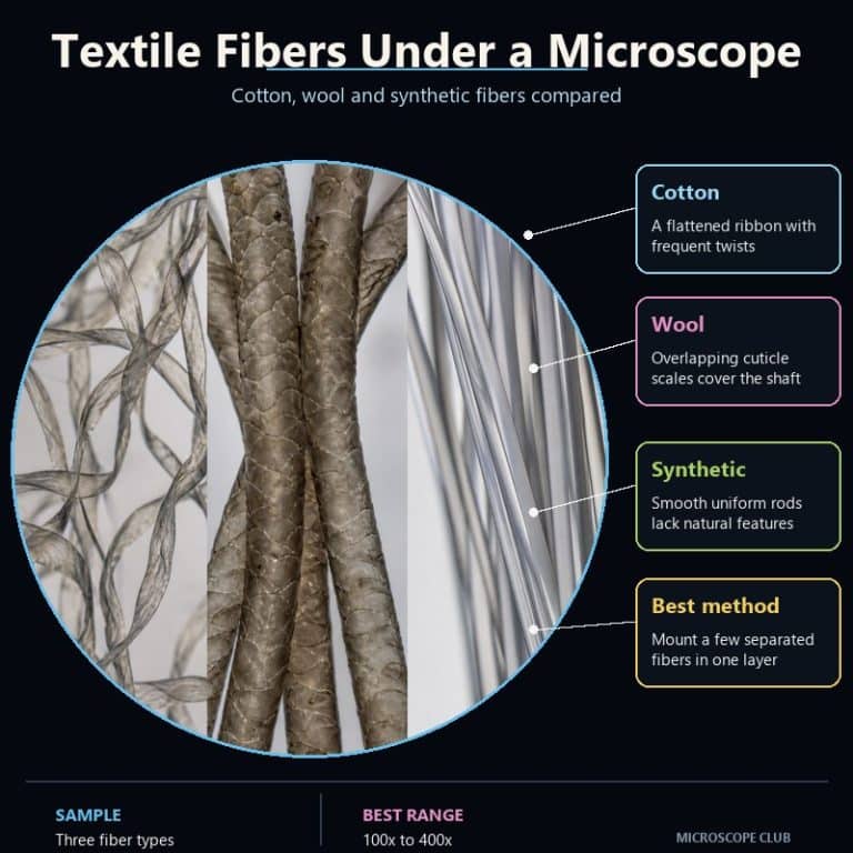
Textile fibers under a microscope give up their identity in seconds, long before a chemical test is needed. Cotton shows up as a flat, twisted ribbon; wool as a cylinder wrapped in overlapping scales; and synthetic fibers like nylon and polyester as a smooth, featureless rod. Once you know these three shapes, you can pull a single thread from almost any fabric and name it the way a forensic fiber examiner would — by structure, not color.
How to Prepare a Fiber Sample
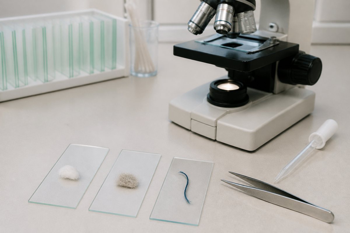
Getting a usable view starts before the slide ever touches the stage. A whole thread or piece of yarn is a bundle of dozens of fibers twisted together, and under a microscope that bundle turns into an unreadable black tangle. The fix is simple but easy to skip: tease out one single fiber with a pair of fine tweezers before you mount anything.
- Pull a loose fiber from a frayed edge or seam allowance so you don’t damage the garment.
- Lay the single fiber straight across the center of a clean glass slide.
- Add one drop of water directly over the fiber — a dry mount traps air bubbles and glare.
- Lower the coverslip at an angle rather than dropping it flat, so it doesn’t push the fiber into a curl.
- Start at 100x total magnification to find and straighten the fiber, then move up to 400x to study scales or twists.
Light control matters as much as the mount. These fibers are nearly transparent, so flooding them with light washes out the very details you’re trying to see. Close the condenser diaphragm down for contrast — the opposite of what you’d do for a stained slide. A drop of water is usually enough, though glycerin or mineral oil gives even better contrast if you have it on hand. For a full walkthrough of the mounting steps themselves, see our guide to making a wet mount slide.
Cotton Under a Microscope — the Twisted Ribbon
Cotton fiber is almost pure cellulose, and its shape under the lens looks like a wrung-out garden hose or a twisted party streamer: a flat, translucent ribbon that spirals along its length and reverses the direction of its twist as your eye travels down it. Each of those spiraling twists is called a convolution, and a single fiber can carry roughly sixty of them per centimeter, according to fiber structure research summarized by ScienceDirect.
The convolutions exist because a growing cotton fiber starts as a round, fluid-filled tube with a hollow central canal called the lumen. As the boll opens and the fiber dries, that canal collapses inward, flattening the round tube into a spiraled ribbon. In cross-section this leaves cotton with its signature kidney-bean shape, with the collapsed lumen still visible as a thin dark line running down the center. Diameters typically run 12–20 µm — finer than a human hair, which averages 50–100 µm.
Watch for one common trap: mercerized cotton, which has been treated with caustic soda to boost strength and dye uptake, swells and loses most of its twists. It looks rounder and smoother than raw cotton and gets misfiled as a synthetic more often than any other natural fiber. The tell is still there if you look for it — check for the faint central lumen running the length of the fiber, even on a mercerized sample that looks deceptively smooth.
Wool Under a Microscope — the Scaled Cylinder
Wool fiber is a protein — keratin, the same material as hair and fingernails — and it announces itself with a texture cotton and synthetics simply don’t have. Overlapping cuticle scales run up the shaft like stacked cups, pinecone scales, or the plates on a pinecone, all pointing toward the fiber’s tip. At 400x, the edges of those scales catch the light as faint stepped lines, and the whole fiber reads as a thicker, more solid rope compared to cotton’s flat ribbon.

Those scales aren’t just a visual signature — they’re the reason wool felts and shrinks. Under heat, moisture, and agitation, the raised scale edges interlock in one direction and resist sliding back, a directional-friction effect that pulls the fibers tighter together with every wash cycle. Research on wool finishing confirms this mechanism directly: treatments that strip or smooth the scale layer measurably reduce felting, while wool with its scale structure intact remains prone to shrinkage under mechanical action in a humid, hot environment.
Coarser wool sometimes shows a medulla, a hollow core running through the center of the fiber, while fine wool like merino often shows none. Diameters range from about 15 µm in fine merino up to 40 µm in coarse wool, and a gentle natural waviness called crimp is visible even at low magnification.
Synthetic Fibers Under a Microscope — the Smooth Rod
Nylon and polyester look almost nothing like cotton or wool: a dead-smooth, uniform cylinder with no twists and no scales, unnervingly consistent in width from end to end — think a glass rod or a strand of clear, cooked spaghetti. That uniformity isn’t an accident of nature; it’s a direct result of how the fiber is made. Molten or dissolved polymer is forced through a spinneret, a metal plate studded with tiny holes, and every fiber that emerges takes on the exact, repeatable diameter of the hole it passed through.

Look closely and you may spot tiny dark specks suspended inside the otherwise clear fiber, like flecks of black pepper. That’s delustrant — usually titanium dioxide — added during manufacturing specifically to cut the fiber’s natural shine. As reference sources on textile delustrants explain, powdered titanium dioxide gets mixed into the polymer before spinning, and manufacturers dial the amount up or down depending on whether they want a bright, semi-dull, or fully matte finish. Natural fibers don’t have anything like it, which makes delustrant specks one of the more reliable synthetic tells once you know to look for them.
Cross-sections are usually round, though manufacturers also engineer trilobal (three-lobed) shapes for added sparkle in carpet fibers, and diameters run anywhere from roughly 10 to 50 µm depending on the fiber’s denier.
Cotton vs Wool vs Synthetic: The Identification Table
Side by side, the three fiber families separate cleanly on five points: how they look lengthwise, their cross-section, their diameter, their surface, and the one feature that gives each one away.
| Fiber | Longitudinal view | Cross-section | Diameter | Distinguishing feature |
|---|---|---|---|---|
| Cotton | Flat, twisted ribbon (convolutions) | Kidney-bean, with lumen | ~12–20 µm | Reversing twists + central lumen |
| Wool | Cylinder with overlapping scales | Round to oval; medulla in coarse wool | ~15–40 µm | Shingle-like cuticle scales |
| Nylon / Polyester | Smooth, uniform rod | Round (sometimes trilobal) | Uniform, ~10–50 µm | Featureless uniformity; delustrant specks |
In practice, work through the table in order: first check for scales — if they’re there, it’s wool, no further test needed. If there are no scales, look for twists and a central lumen; a ribbon shape with visible convolutions is cotton, even if it’s been mercerized smooth. If the fiber shows neither scales nor twists — just a flat, dead-uniform rod — you’re looking at a synthetic, and delustrant flecks (if present) confirm it. Color never enters the decision; dyes sit on the surface and tell you nothing about the fiber underneath.
This shape-first approach is exactly how forensic examiners work a real fiber comparison. Crime labs identify unknown fibers by structure under comparison and polarized-light microscopy well before any chemical test, and forensic fiber research from the National Institute of Justice shows just how much identifying detail a single fiber can hold under the right microscope setup.
Seeing the three shapes side by side on video makes the differences click faster than any description. This comparison from Microbehunter Microscopy walks through natural and synthetic fibers under magnification:
Beyond the Big Three — Linen and Silk at a Glance
Two more fibers are worth a quick look if you want to round out your identification skills. Linen, spun from the flax plant, is cellulose like cotton but shows a completely different tell: distinct node markings that cross the fiber at intervals, giving it a bamboo-jointed look. Its cross-section is polygonal rather than kidney-shaped, though it does share cotton’s central lumen.
Silk is a protein fiber, like wool, but spun by silkworms rather than sheared from an animal. Under the microscope it’s fine, smooth, and only slightly irregular — smooth enough to be mistaken for a synthetic at first glance. The giveaway is its cross-section: silk is triangular, which is what bends light and gives silk fabric its natural sheen, whereas a true synthetic stays perfectly round or trilobal and shows no natural irregularity at all.
Frequently Asked Questions
What magnification do you need to see fabric fibers?
Most textile fiber features are visible starting around 100x, which is enough to spot cotton’s twists or wool’s general scale pattern. Push to 400x to resolve fine detail like individual scale edges or delustrant specks in synthetics.
Can you tell silk from synthetic under a microscope?
Yes, though it takes a closer look than cotton or wool. Silk has a triangular cross-section and slight natural irregularity along its length, while true synthetics stay perfectly uniform in diameter and typically show a round or trilobal cross-section.
Why does wool shrink or felt?
Wool’s overlapping cuticle scales interlock under heat, moisture, and agitation. The scales catch and hold in one direction, pulling fibers tighter together with each wash — the same scale structure that identifies wool under the microscope is what causes the shrinkage.
Why does my fiber sample look like a black tangled mess instead of a single shape?
You’re likely viewing a whole thread or yarn rather than one fiber. Threads are bundles of many fibers twisted together, and under magnification that bundle overlaps into an unreadable tangle. Tease out and straighten a single fiber with tweezers before mounting.
Does the color of a fabric help identify the fiber?
No — dyes sit on the fiber’s surface and can be applied to almost any fiber type, so color tells you nothing about what’s underneath. Identification relies entirely on structure: the presence or absence of twists, scales, or uniform smoothness.
Can I use a household water drop instead of a proper mounting medium?
A single drop of water works fine for a quick look and is what most of the observations in this guide are based on. Glycerin or mineral oil will give slightly better contrast if you have either on hand, but they aren’t required to see the basic shapes.
Why does mercerized cotton look so different from raw cotton?
Mercerization treats cotton with caustic soda, which swells the fiber and irons out most of its natural twists, leaving it rounder and smoother. It’s a common misidentification trap — check for the faint central lumen, which survives mercerization even when the twists don’t.
Conclusion
Once you’ve seen cotton’s twisted ribbon, wool’s shingled scales, and synthetic’s dead-uniform rod side by side, you stop needing a label to know what you’re looking at. The shape-first method here — scales mean wool, twists and a lumen mean cotton, featureless uniformity means synthetic — works on everything from a lab sample to a mystery scrap pulled from your own closet, the same way it works in the fiber web inside a sheet of paper or on a strand of human hair under a microscope, which shares wool’s cuticle-scale structure.
Have you pulled a fiber from an old shirt or a ball of yarn and checked it under your own scope? Tell us what you found in the comments below — and if you’re still getting comfortable with the prep work, our guide to making a wet mount slide covers the technique used throughout this article in more depth.