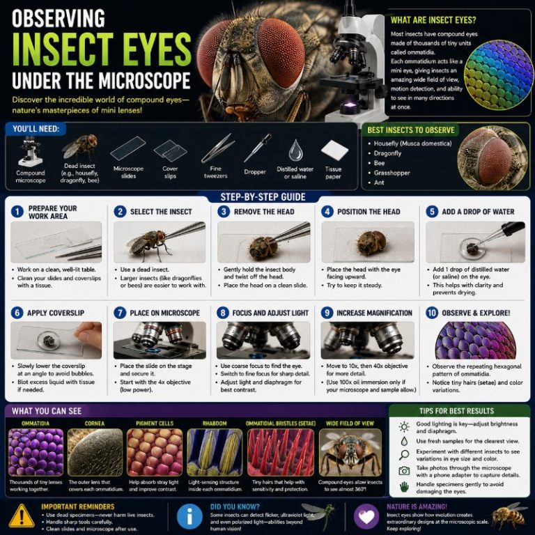
Viewing insect eyes under the microscope reveals one of nature’s most striking optical structures: a dense honeycomb of hexagonal facets, each one a separate lens called an ommatidium. At 20–40x on a stereo microscope, the mosaic array snaps into sharp focus — and if you rotate the specimen, a dark spot appears to track your eye. That’s the pseudopupil, and it’s just one of the surprising details waiting when you look closely at an insect’s eye.
What Do Insect Eyes Look Like Under a Microscope?
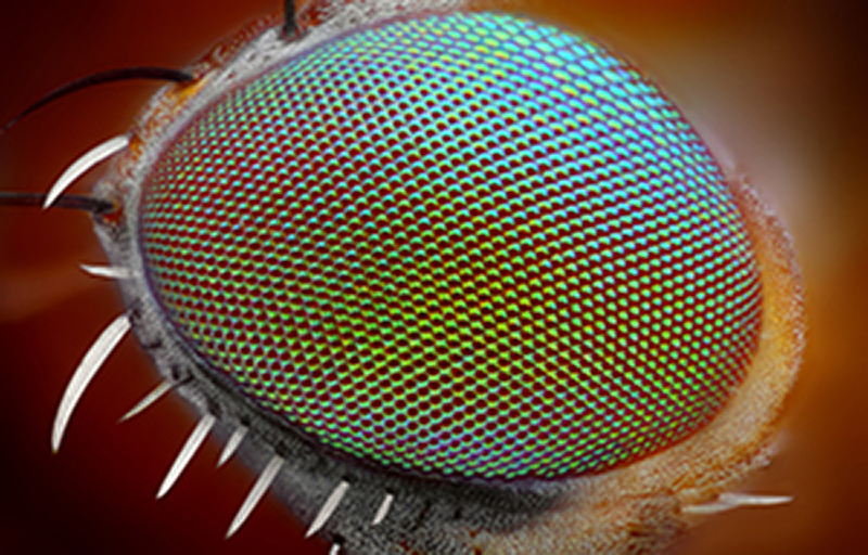
A compound eye looks like a soccer ball made of tiny hexagonal tiles. Each tile is the outer corneal lens of one ommatidium — an independent optical unit with its own lens, light-focusing cone, and photoreceptor cells underneath. Most adult insects carry two large compound eyes, one on each side of the head, plus (in many species) three small simple eyes called ocelli on top of the head. Don’t confuse the ocelli with ommatidia — they detect light intensity rather than form images.
Under a stereo microscope at 20–40x, the honeycomb facet array is unmistakable. The surface shimmers with structural color that shifts as you change the light angle — caused by microstructure interference, not pigment. At higher compound-microscope magnifications (100–400x), you can resolve fine texture between facets, tiny sensory hairs, and on prepared sections, the internal anatomy of individual ommatidia.
Compound Eyes vs. Human Eyes
What Makes a Compound Eye Different
A human eye uses a single large lens that projects a focused image onto the retina — much like a camera. A compound eye uses hundreds to tens of thousands of tiny lenses, each pointed in a slightly different direction. There is no single focal plane; instead, the brain assembles a coarse mosaic from signals of all ommatidia simultaneously. The result is not a sharp photograph but a wide-angle, motion-sensitive image — excellent for detecting fast movement across a broad field of view.
| Feature | Compound Eye (insect) | Simple Eye (human) |
|---|---|---|
| Number of lenses | Hundreds to ~30,000 | 1 |
| Image type | Coarse mosaic | High-resolution focused image |
| Spatial resolution | Low | High |
| Motion detection | Excellent (high flicker-fusion rate) | Good, but slower |
| Field of view | Wide (often near 360°) | ~180° total |
| UV sensitivity | Many species (bees, flies) | No |
| Polarized light | Yes (many species) | No |
The Structure of an Ommatidium
What Each Facet Contains
Each ommatidium is roughly 10–35 micrometers (µm) across — small enough that the whole eye looks smooth to the naked eye. It’s a self-contained optical unit stacked from front to back. For a detailed overview of these optics, see Arizona State University’s compound eye explainer.
- Corneal lens — the hexagonal outer facet visible on the eye surface; refracts incoming light downward.
- Crystalline cone — a transparent cone beneath the corneal lens that focuses and funnels light.
- Retinula cells — typically eight photoreceptor cells arranged around a central light-guiding structure called the rhabdom; they convert light into nerve signals.
- Pigment cells — insulating cells that wrap the ommatidium to prevent light bleeding into neighboring units (in apposition eyes).
Why Facets Are Hexagonal
Hexagonal packing is the most efficient geometry for covering a curved surface — it maximizes the number of lenses per unit area while minimizing gaps. You see the same pattern in honeycombs and bubble rafts. A few insect families show square or rounded facets, but hexagonal is the canonical form you’ll encounter at the eyepiece.

Apposition vs. Superposition Eyes
Not all compound eyes work the same way:
- Apposition eyes (day-active insects: houseflies, bees, dragonflies) — each ommatidium is optically isolated from its neighbors, giving better spatial discrimination in bright light. Under the microscope these eyes typically appear dark or matte.
- Superposition eyes (night or crepuscular insects: most moths, many beetles) — optical isolators are absent or reduced, allowing light from many facets to pool onto one receptor for greater sensitivity in low light. Many have a reflective layer (tapetum) that makes the eye glow greenish-gold in reflected light — the same mechanism as eyeshine in cats.
How Many Lenses? Ommatidia Counts by Insect
The number of ommatidia varies enormously between species and reflects each insect’s lifestyle. Dragonflies, aerial predators that catch prey in flight, have the highest counts:
| Insect | Ommatidia per eye (approx.) |
|---|---|
| Dragonfly | Up to ~28,000–30,000 |
| Honey bee (worker) | ~4,000–5,000 |
| Housefly | ~3,000–6,000 |
| Butterfly / moth | Several thousand (varies widely) |
| Worker ant | ~100–1,000 (species dependent) |
More ommatidia does not work like megapixels on a camera — even a dragonfly’s 30,000-facet eye produces a coarser image than a human eye. The advantage is wide field of view and extreme motion sensitivity, not fine detail.

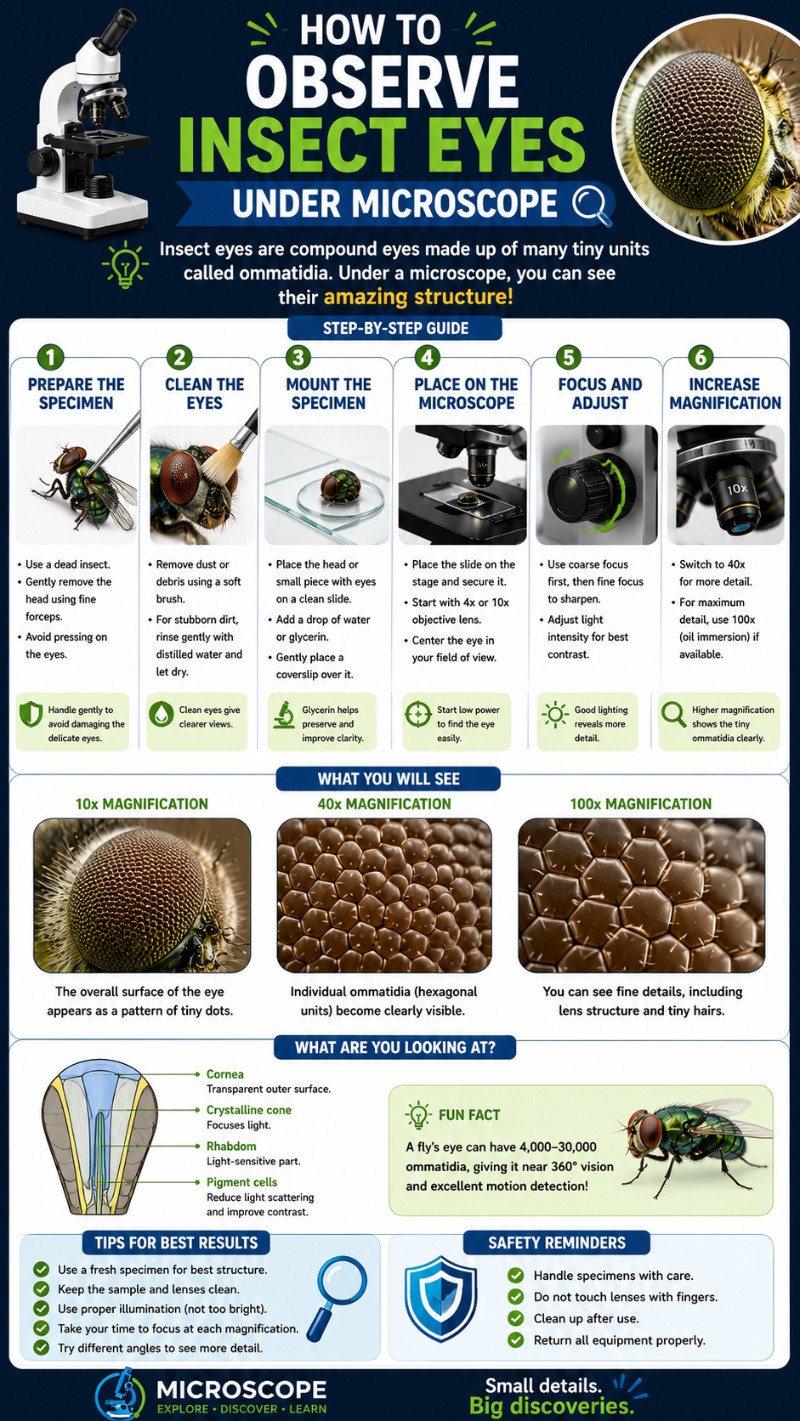
What You’ll Actually See — Magnification by Magnification
Understanding magnification vs. resolution before you start will help you choose the right setting for each stage of observation.
| Magnification | What’s visible |
|---|---|
| Naked eye / hand lens (2–5x) | Smooth domed surface; overall shape and color |
| Stereo microscope 10–20x | Facet array begins to resolve on larger insects; iridescent color bands become clear |
| Stereo microscope 20–40x | Individual hexagonal facets clearly visible; pseudopupil appears as a dark spot that moves as you rotate the specimen |
| Compound microscope 40–100x | Individual ommatidia sharply resolved; sensory hairs between facets; structural color variation across the array |
| Compound microscope 400x | Fine surface texture; on a thin section, internal ommatidium anatomy (corneal lens outline, cone shadow) |
The Pseudopupil — What Is It?
When you look at a compound eye at 20–40x and slowly rotate the specimen, a dark spot moves with your viewing angle, always appearing to look back at you. This is the pseudopupil. It is not a real pupil — insects cannot dilate or constrict it. It appears because the ommatidia pointing directly at your eye absorb incoming light rather than reflecting it, making that cluster appear black against surrounding facets. Rotate the specimen 30 degrees and the pseudopupil shifts to the ommatidia now aimed at you. It is a reliable indicator that your specimen is correctly oriented and your lighting is working.
Best Microscope for Viewing Insect Eyes
Stereo (Dissecting) Microscope — the Recommended Tool
A stereo microscope is the right choice for viewing a whole insect eye. It provides reflected light from above, gives a true 3D upright view, and operates at the 10–40x range where the facet array is most impressive. You don’t need to section or flatten the specimen — just position the head or eye on the stage and focus. For background on stereo microscope optics and setup, Nikon MicroscopyU is an excellent free reference.
Compound Light Microscope — for Sections and Higher Detail
A compound light microscope uses transmitted light, which won’t work well on an intact, opaque, curved eye. It excels with prepared thin sections or squash mounts, where you can use objective lenses at 40–400x to resolve internal ommatidium structure. If you want this level of detail without doing your own sectioning, buy a commercially prepared insect eye slide — they are inexpensive and give clean results.
USB/Digital and Pocket Microscopes for Beginners
A USB or digital microscope is a solid budget entry point for capturing images of the facet array at low magnification. Many models reach 40–100x and can produce good photographs of a dragonfly or housefly eye without much investment. See the full range of types of microscopes to find the right fit for your budget and goals.
How to Prepare an Insect Eye Specimen
Sourcing Your Specimen
You don’t need to collect live insects — windowsill casualties work fine. Good candidates:
- Houseflies — easy to find, good facet count, eyes large enough for a first observation.
- Dragonflies — large compound eyes that nearly wrap around the head; ideal for seeing the pseudopupil and structural color.
- Honey bees or bumblebees — often found dead near flowers or on windowsills; clear hexagonal facets.
- Ants — worker ants have small eyes with few facets, but they make an interesting size comparison. See our guide on ants under the microscope for setup tips.
Fresh vs. dried: Fresh specimens retain shape and iridescent color best, but the eye dries and clouds within a few hours. Dried specimens are easier to handle but facets can collapse or shrink, and the surface may look dusty. For a first observation, a fly or bee that died within the last day or two works well.
Mounting for Stereo Microscope Viewing
- Place the dead insect or detached head on a piece of modeling clay (plasticine) or double-sided tape on a glass slide.
- Orient the eye to face upward toward the objective lens.
- Use the stereo scope’s reflected (incident) top light angled slightly from one side — not flat overhead. Angled oblique lighting makes facet relief pop and reveals the iridescence. Flat overhead light flattens the hexagonal array.
- Start at low magnification (10x), locate the eye surface, then zoom to 20–40x.
- Slowly rotate the specimen and watch the pseudopupil shift position.
Prepared Slides for Compound Microscope Viewing
For transmitted-light compound microscope work, a thin section is required — the intact eye is too opaque and thick. The easiest route is a commercially prepared insect compound eye slide, available from biological supply companies for a few dollars. These show internal ommatidium structure at 100–400x without any sectioning skill on your part. If you prefer to make your own, follow the steps in our guide on how to prepare microscope slides and use a very thin razor section through the eye, mounted in glycerin.
What Insects Actually See Through These Eyes
A common misconception is that insects see thousands of tiny copies of the same image, one through each lens. They don’t. Each ommatidium captures a single “pixel” of light from its narrow acceptance angle, and the brain assembles all the pixels into one combined mosaic image — coarse, wide-angle, and optimized for motion detection rather than fine detail.
The tradeoffs are real: a fly cannot read a newspaper, but it can detect a hand moving toward it from almost any direction with extraordinary speed (the neural reason why a fly is so hard to swat). Many species also see into the ultraviolet range and use polarized light for navigation. The compound eye is not a worse camera — it’s a different instrument optimized for a very different lifestyle. For a deeper look at the biology, see Encyclopædia Britannica’s compound eye article.
Frequently Asked Questions
How much does it cost to set up a microscope for viewing insect eyes?
You can start cheaply: a handheld USB or pocket digital microscope (40–100x) runs roughly $20–$50 and is enough to capture a fly or dragonfly facet array. A basic entry-level stereo (dissecting) microscope — the better tool for whole intact eyes — starts around $80–$150, while quality hobby models with good optics and lighting run $200–$400. Commercially prepared insect eye slides for a compound scope cost only a few dollars each, so you don’t need to spend much to get a clear first view.
How do you photograph an insect eye through a microscope?
The simplest method is a smartphone adapter that clamps your phone’s camera over the eyepiece — alignment is critical, so a model that lets you center and tilt the camera precisely gives the best results. Because high magnification has a very shallow depth of field, a single shot rarely keeps the whole curved eye sharp; photographers fix this with focus stacking, taking a series of images at slightly different focus depths and merging them in software like Helicon Focus. Use angled (oblique) lighting to bring out the facet relief and iridescence, and shoot at the lowest magnification that fills the frame.
How long do insect specimens stay viewable, and how do you store them?
A fresh specimen keeps its shape and iridescent color best but the eye dries and clouds within a few hours, so observe it the same day. Dried specimens last for years if stored correctly: keep them out of direct light in a low-humidity environment (around 20°C and 50% relative humidity is ideal) and avoid sealed airtight containers, which can trap moisture and grow mold. The main long-term threat is pests like carpet beetles (Dermestidae), so freeze new specimens for a few days to kill any hidden larvae and check stored collections every few months.
What are the most common mistakes beginners make when viewing insect eyes?
The biggest one is using flat overhead lighting, which washes out the hexagonal facets — use angled (oblique) top light instead to reveal relief and structural color. Beginners also tend to jump straight to high magnification; always start at the lowest power (10x), find and focus the eye, then zoom in. Other frequent errors are letting a fresh eye dry out before viewing, and reaching for a compound microscope on an intact opaque eye when a stereo microscope with reflected light is the right tool for a whole specimen.
Conclusion
Insect eyes are one of the most rewarding specimens in hobby microscopy — compact, easy to find, and visually spectacular at even modest magnification. The hexagonal facet mosaic, the iridescent structural color, and the tracking pseudopupil are all visible on a windowsill fly under a basic stereo scope at 20–40x. Go deeper with a prepared slide and a compound microscope and you’ll see the ommatidium anatomy that took scientists decades to fully map.
Have you tried viewing insect eyes at home? Drop your results — or your questions — in the comments below. Which insect gave you the clearest view of the facet array?
Originally posted 2026-06-13 14:07:06.