Spirogyra, a filamentous freshwater green alga, reveals one of the most visually striking structures you will encounter under a light microscope: a continuous ribbon of vivid green chloroplast wound in a perfect helix along every cell. Pull a pinch of pond-scum mat from the water in spring, mount a small piece on a slide, and at 100× that spiral snaps into focus in a way that makes people stop and look twice.
What Is Spirogyra?
Spirogyra is an unbranched filamentous green alga belonging to the family Zygnemataceae. Each filament is a chain of identical cylindrical cells joined end-to-end at flat cross-walls called septa. The defining feature — the one no other common freshwater alga shares in quite the same way — is one to sixteen ribbon-like chloroplasts arranged in a spiral or helical pattern inside each cell.
Despite being nicknamed “pond scum,” Spirogyra is one of the most common and widely distributed freshwater green algae on Earth, found globally in eutrophic freshwater habitats.1 It thrives in eutrophic ponds, ditches, lake margins, and slow-moving streams, and it is most abundant from spring into early summer. The genus is large — several hundred species are recognised, with estimates commonly around 400 — but identifying individual species requires examining reproductive structures, particularly zygospore morphology, which are rarely present in a casual collection.2
Other informal names — water silk and mermaid’s tresses — come from its feel: pull a floating mat out of the water and it slides through your fingers with a slippery, almost silky texture, nothing like the gritty feel of sand or the rough texture of dried grass. That slipperiness comes from a mucilage sheath surrounding each filament.3
The Structure of Spirogyra

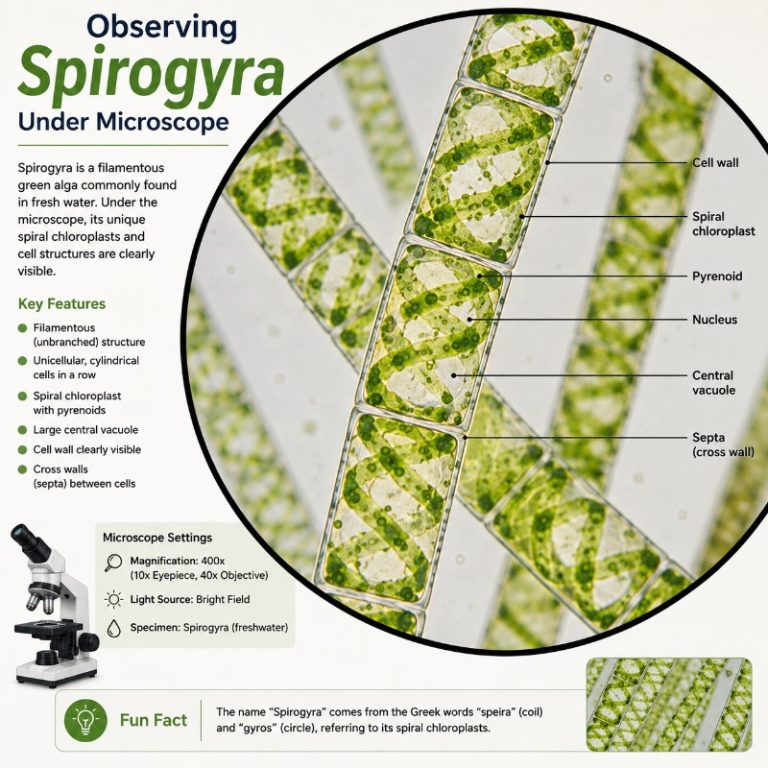
Every component of Spirogyra’s structure is visible — to varying degrees — with a good compound light microscope:
- Cell wall — A two-layer wall of cellulose fibers provides structural rigidity and turgor support. The outer layer merges into the mucilage sheath.
- Mucilage sheath — The slippery coating around each filament. It aids buoyancy, substrate contact, and protection.
- Chloroplast — The star of the show. Each cell contains one to sixteen ribbon-like chloroplasts arranged in a spiral or helix. This is the defining visual feature of Spirogyra and is clearly visible at 100×.
- Pyrenoids — Bead-like protein bodies embedded at regular intervals along each chloroplast ribbon. They are the site of a carbon-concentrating mechanism (CCM) that concentrates CO₂ around RuBisCO to boost carbon fixation, and they are the focal point for starch storage — starch plates form around them. At 400× they appear as a row of small bright dots along the spiral ribbon.
- Large central vacuole — A single large vacuole occupies most of the cell’s interior, pushing the cytoplasm, chloroplasts, and nucleus to the periphery or suspending the nucleus centrally on cytoplasmic strands. No specific percentage should be assumed; it is simply the dominant internal space.
- Nucleus — Sits suspended in the centre of the cell, held by cytoplasmic strands radiating from the cell periphery. Visible as a slightly denser, rounded body at 400×.
- Cytoplasmic strands — Fine threads of cytoplasm that connect the peripheral layer to the centrally-suspended nucleus, crossing the vacuole.
- Cytoplasm — Confined to a thin peripheral layer and the strands around the nucleus, due to the large vacuole.
Gas exchange note: Spirogyra has no stomata. Stomata are structures found only in land plants and some bryophytes. Spirogyra is an alga; every cell is in direct contact with the surrounding water, and CO₂ and O₂ move by simple diffusion across the cell wall and membrane into the water column. The oxygen produced during photosynthesis is also what causes Spirogyra mats to become buoyant and float to the surface as they mature.
Where to Find and Collect Spirogyra
The best time to collect Spirogyra is spring through early summer, when growth peaks and the algae are at their most photosynthetically active. Look for bright-green, floating mats in still or slow-moving eutrophic water — garden ponds, roadside ditches, shallow lake margins, or calm backwaters of streams. From a few feet away the mat looks like a fuzzy green blanket on the water surface. Up close it is loose and airy, not compacted.

The sensory test is reliable: scoop a handful and let it slide between two fingers. Spirogyra feels slippery and silky — the mucilage sheath is distinctly different from the gritty texture of diatom mats or the rough feel of Cladophora. If it feels silky, it is very likely Spirogyra or one of its close relatives (Zygnema or Mougeotia — see the ID table below).
To collect a sample, use a small jar or a zip-lock bag, scoop up a small piece of the mat along with some of the surrounding water, and seal it. Keep it cool and out of direct sunlight during transport. A pea-sized clump is more than enough for dozens of slides; you do not need much.
How to Observe Spirogyra Under the Microscope

A standard compound light microscope with 40×, 100×, and 400× total magnification covers everything Spirogyra has to show. Total magnification equals eyepiece magnification multiplied by objective magnification — so a 10× eyepiece with a 10× objective gives 100× total. Follow this step-by-step protocol:

- Prepare the slide. Place a single drop of the pond water containing the mat on a clean glass slide. Using tweezers or a dissecting needle, tease out a small piece of filament — a few millimetres long. Spirogyra filaments can be several centimetres; if they are too long they will bunch under the coverslip and become unreadable. Trim to size. Lower a coverslip at an angle to avoid trapping large air bubbles. What you should see at this point with the naked eye: a faint green thread in clear water.
- Scan at 40× total (4× objective + 10× eyepiece). Locate a filament and centre it in the field of view. What you see: long, straight, parallel green threads. The spiral pattern is not yet visible — cells look like green tubes joined end to end. Use this magnification to find a clean section of filament with no overlapping strands.
- Switch to 100× total (10× objective). Refocus carefully. What you see: this is the payoff view. The spiral or helical ribbon chloroplast inside each cell becomes clearly visible — a continuous green band winding in a steady corkscrew from one end of the cell to the other. The cross-walls (septa) between cells are sharp and straight. The central vacuole appears as a lighter, less-dense region inside each cell. This is what people mean when they describe Spirogyra as beautiful under the microscope.
- Switch to 400× total (40× objective). What you see: individual features within the cells resolve. The pyrenoids appear as a row of small bright or slightly greenish-white bead-like dots along the ribbon of chloroplast. The nucleus becomes visible as a dense oval body suspended in the centre of the cell, held by fine cytoplasmic strands crossing the vacuole. The cell wall is crisp and double-layered. Reduce your light intensity slightly at this magnification — too much illumination bleaches the green and washes out the detail.
- Optional: iodine stain for pyrenoids. Add a small drop of iodine solution (I₂KI) to the edge of the coverslip and allow it to diffuse under. The starch plates around pyrenoids stain blue-black, making each bead along the chloroplast ribbon pop with high contrast. The spiral chloroplast itself is already vivid green and needs no stain to see; iodine is purely for showing starch localisation around pyrenoids.
- Optional: methylene blue for nuclear contrast. A dilute drop of methylene blue improves contrast on the nucleus and cytoplasm, making the centrally-suspended nucleus easier to locate. Use sparingly — a little goes a long way.
Common beginner problems
The most frequent mistake is putting too much material on the slide. A thick clump of filaments overlaps into an unreadable green mass; you need a single layer of a few strands. The second common error is coverslip pressure: pressing the coverslip down bursts the cells and you will see only green smears. Lower it gently and let capillary action do the work. Over-illumination is the third problem — at 400× especially, high light intensity washes out the chloroplast colour and makes pyrenoids nearly invisible. Turn the condenser aperture down slightly until the green deepens.
How Does Spirogyra Reproduce?

Spirogyra reproduces in three modes: vegetative, asexual (spore-based), and sexual (conjugation).
Vegetative reproduction — fragmentation
The most common mode. Any mechanical disturbance — water turbulence, grazing by invertebrates, even the death of a cell within a filament — can break a strand into fragments. Each fragment regenerates a complete filament. This is how Spirogyra spreads rapidly across a pond surface without any reproductive structures at all.
Asexual reproduction — three spore types
Under adverse conditions — nutrient depletion, desiccation, cold — Spirogyra can produce three types of asexual spores:
- Akinetes — thick-walled resting cells formed when conditions become harsh. The wall (cellulose and pectin) protects against desiccation and freezing. When conditions improve, akinetes germinate into new filaments.
- Aplanospores — thin-walled, non-motile spores. The cell protoplast contracts, walls off, and is eventually released when the parent filament dies. They germinate when conditions become favourable again.
- Azygospores (parthenospores) — formed when gametes fail to fuse properly during a conjugation attempt and instead develop into spores independently.
Note: zygospores are the product of sexual reproduction (conjugation), not asexual — these two categories must not be confused.

Sexual reproduction — conjugation
Conjugation is the event that makes Spirogyra genuinely exciting to watch under the microscope, and it is observable with a standard light microscope if you are lucky enough to collect material at the right time.
Scalariform (ladder) conjugation occurs between two separate filaments that come to lie parallel to each other. Corresponding cells in each filament push out short tubes (papillae) toward each other; the tubes meet and fuse, forming a channel. The contents of one cell migrate through this tube — not by swimming with a flagellum (Spirogyra gametes are non-flagellate) but by an amoeboid-like movement — and fuse with the contents of the other cell. The result is a thick-walled zygospore. Seen at 100×, two filaments connected by a row of these bridges look unmistakably like a ladder, which is how the process got its name.
Lateral conjugation occurs between adjacent cells of the same filament, without involving a second filament. A tube forms between one cell and its neighbour; the gamete migrates and a zygospore forms within the filament.
Zygospores are thick-walled, dark, and oval or lens-shaped — very different in appearance from a vegetative cell. Species identification in Spirogyra relies heavily on zygospore morphology, which is why it is difficult to identify species from vegetative material alone.4
What Does Spirogyra Eat? (Energy Source)

Spirogyra is a photoautotroph — it produces its own energy through oxygenic photosynthesis, using chlorophyll in the spiral ribbon chloroplasts to absorb light energy and convert CO₂ and water into carbohydrates. Chlorophyll absorbs red and blue wavelengths and reflects green, which is why Spirogyra has its characteristic vivid green colour.
Gas exchange is by direct diffusion across the cell surface into the surrounding water — there are no stomata, no specialised pores of any kind. The oxygen generated during photosynthesis dissolves into the surrounding water or forms tiny bubbles that accumulate under the mat. As bubbles grow, they lift the mat toward the surface — a Spirogyra bloom rising to the top of a pond is simply oxygen production becoming visible.
Pyrenoids along the chloroplast ribbon are the hubs of carbon fixation. They run a carbon-concentrating mechanism that pumps CO₂ around the enzyme RuBisCO, improving fixation efficiency. Starch accumulates in plates around each pyrenoid, which is why an iodine stain turns those locations blue-black.
How to Identify Spirogyra vs Similar Algae
Several filamentous green algae can be collected from the same habitat as Spirogyra, and at first glance a green thread is a green thread. The chloroplast shape is the single most reliable feature visible under a light microscope to distinguish the three most common look-alikes. Use the table below at 100× total magnification — that is the magnification at which chloroplast shape is clearest. Readers who have also looked at plant cells under the microscope will notice immediately that Spirogyra’s chloroplasts are far more organised and visually distinct than those found in land-plant leaf sections.
| Alga | Chloroplast shape (at 100×) | Number per cell | Quick ID note |
|---|---|---|---|
| Spirogyra | Ribbon-like spiral / helix | 1–16 | Unmistakable corkscrew; pyrenoids visible as beads along the ribbon |
| Zygnema | Two star-shaped (stellate) bodies, one at each end of the cell | 2 per cell (always) | Looks like two green asterisks per cell; no spiral |
| Mougeotia | Single flat axial plate (often twisted to face the light) | 1 | Ribbon lying flat or edge-on depending on light angle; may twist as you watch |
If you can see the spiral, you have Spirogyra. If you see two star shapes, it is Zygnema. If you see a flat plate that may twist, it is Mougeotia. All three are in the same family (Zygnemataceae) and often occur together in the same mat, so knowing this table lets you confirm your identification on the spot rather than guessing.5
What Are the Types of Spirogyra?
Spirogyra is a large genus with several hundred species — commonly around 400, with some taxonomic treatments listing 350–500 or more. Visually distinguishing one species from another is difficult from vegetative material alone, because the characters that separate species — zygospore shape, wall ornamentation, size, and the specifics of the reproductive structures — are only present during conjugation. Two filaments sitting side-by-side in a mat may belong to different species and be indistinguishable without reproductive material. This is why collecting during the conjugation season (spring, coinciding with peak growth) gives the best chance of finding identifiable material.
Frequently Asked Questions
Is Spirogyra a plant or an alga? Does it have stomata?
Spirogyra is an alga, not a plant. It has no stomata — stomata are specialised pores found only in land plants (and some bryophytes). Because every Spirogyra cell is in direct contact with the surrounding water, gas exchange happens by simple diffusion across the cell surface. No pores, no opening and closing, no guard cells.
Why is Spirogyra called pond scum?
The name refers to the way Spirogyra mats accumulate on the surface of ponds and slow water as the algae mature and their photosynthetic oxygen production lifts them up. Up close the mat is loose, green, and slimy — hence “scum.” The alternative name “water silk” is arguably more accurate: the mucilage sheath gives the filaments a genuinely silky feel between the fingers.
What magnification do you need to see Spirogyra’s chloroplast?
The spiral chloroplast becomes clearly visible at 100× total magnification (10× eyepiece × 10× objective). At 40× you can see the filaments as green threads but the spiral is not resolved. At 400× you resolve pyrenoids, the nucleus, and cross-walls. You do not need an electron microscope — a standard student compound microscope handles everything Spirogyra offers.
What does Spirogyra look like under a microscope?
At 100×: long, parallel chains of rectangular cells, each containing a vivid green ribbon chloroplast that spirals the full length of the cell like a coiled spring. The cells are separated by crisp straight cross-walls. At 400×: bead-like pyrenoids become visible along the chloroplast ribbon, and the nucleus appears as a denser oval body suspended centrally by fine cytoplasmic strands crossing the large vacuole.
How do you collect Spirogyra?
Look for bright-green floating mats in still or slow-moving eutrophic ponds or ditches in spring or early summer. The mat feels slippery and silky (not gritty) between your fingers. Collect a pea-sized piece with some surrounding pond water in a sealed jar. Keep it cool and out of direct sunlight. This small amount is enough for many slides — you do not need a large sample. The same water sample often contains plankton under the microscope worth exploring on the same session.
Conclusion
Spirogyra rewards any microscopist who takes the time to find it: the spiral chloroplast at 100× is one of the most immediately beautiful structures a compound light microscope can show. To get the most out of a slide, collect a small mat sample from a still pond in spring, mount just a few millimetres of filament in a drop of the pond water, and step through 40×, 100×, and 400× in sequence — each magnification adds another layer of detail, from the green threads to the helix to the pyrenoid beads along the ribbon. If you find material in conjugation, the ladder of tubes between two filaments is the bonus observation. Start with a good compound light microscope, learn to prepare a clean wet mount, and Spirogyra will not disappoint. Once you have the technique down, observing yeast under the microscope makes another straightforward next step with the same wet-mount method.
