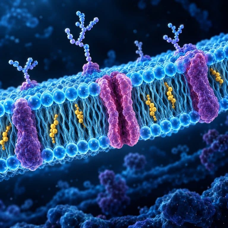
The cell membrane is the thin, selectively permeable phospholipid bilayer that surrounds every cell, controlling which molecules enter and which leave. Built from lipids, proteins, and carbohydrate tags arranged in a constantly shifting pattern called the fluid mosaic model, it holds the cell’s contents in place. It also acts as a gatekeeper between the inside and outside world. Understanding its structure explains everything from why wilted lettuce perks back up in water to how a red blood cell survives in your bloodstream.
What Is the Cell Membrane Made Of?
The cell membrane, also called the plasma membrane, is built around a phospholipid bilayer roughly 7–8 nanometers thick. That’s a number worth sitting with, since a typical school light microscope can’t resolve anything smaller than about 200 nanometers. That gap is the reason you can never actually see the membrane itself under a compound microscope; you only ever see what it does. Cholesterol molecules, embedded proteins, and short carbohydrate chains complete the structure, and each piece has a specific job. For another visual explanation, see OpenStax’s cell membrane components and structure.
The Phospholipid Bilayer
Each phospholipid molecule has a hydrophilic (“water-loving”) phosphate head and two hydrophobic (“water-fearing”) fatty-acid tails. Dropped into water, phospholipids don’t need to be arranged by anything — they spontaneously flip into a bilayer. Heads face outward into the watery cytoplasm and extracellular fluid, while the tails tuck together in the oily middle where water can’t reach them. Scientists call this self-arranging behavior amphipathic, but the plain-language version is simpler: the heads want water, the tails avoid it, and a two-layer sandwich is the only shape that satisfies both at once.
Proteins, Cholesterol, and Carbohydrate Tags
Membrane proteins come in two basic types. Integral (intrinsic) proteins are embedded through the bilayer — many are transmembrane proteins that span it completely and function as channels or carrier/transporter proteins. Peripheral (extrinsic) proteins sit on the surface without crossing the bilayer, usually doing structural or enzymatic work. Cholesterol molecules wedge between the phospholipids in animal-cell membranes and act as a fluidity buffer, keeping the membrane from turning too runny when it’s warm or too stiff when it’s cold. Plant cells rely on related sterols instead. On the outer surface, carbohydrate chains attach to proteins (forming glycoproteins) or to lipids (forming glycolipids) to build the glycocalyx. This coating works like an ID tag, letting the immune system and neighboring cells recognize what they’re looking at.
| Component | What It Is | What It Does |
|---|---|---|
| Phospholipid bilayer | Two layers of phospholipids, heads out, tails in | Forms the membrane’s basic structure and barrier |
| Cholesterol | Steroid molecules wedged between phospholipids | Buffers membrane fluidity across temperature changes |
| Integral proteins | Proteins embedded in or spanning the bilayer | Act as channels and carriers for transport |
| Peripheral proteins | Proteins attached to the membrane surface | Provide structural support and enzyme activity |
| Glycoproteins / glycolipids | Carbohydrate chains on outer-surface proteins and lipids | Form the glycocalyx for cell recognition and signaling |
The Fluid Mosaic Model Explained
The fluid mosaic model is the accepted description of how the cell membrane is put together, proposed by S.J. Singer and Garth Nicolson in 1972. “Fluid” means the membrane isn’t a rigid wall — phospholipids and many proteins drift laterally within their layer the way boats bob and shift on a pond. “Mosaic” describes the look this creates: proteins of different shapes and sizes scattered through the bilayer resemble tiles set into a mosaic floor rather than bricks in a fixed grid. This constant motion matters functionally — it lets proteins cluster where they’re needed and lets the membrane reseal itself after minor damage. It also keeps transport channels mobile instead of locked in one spot.
For a deeper structural breakdown, the National Center for Biotechnology Information’s molecular biology reference walks through the same lipid bilayer chemistry in more technical detail: The Lipid Bilayer.
What Does the Cell Membrane Do? (Functions)
The cell membrane’s core job is acting as a selective barrier: it decides what enters and leaves the cell rather than letting anything through freely. Small nonpolar molecules like oxygen and carbon dioxide slip across on their own, while larger, polar, or charged molecules — glucose, sodium ions, potassium ions — need a protein’s help to cross. Beyond gatekeeping, the membrane gives the cell its physical boundary and shape, and hosts receptor proteins that receive chemical signals from hormones and neighboring cells. It also uses its glycocalyx for cell-to-cell recognition and adhesion. None of these functions work in isolation — a signaling receptor is useless if the barrier around it can’t also hold the cell together. Nature Scitable also provides a useful overview of how cell membranes act as selective boundaries.

How Substances Cross the Membrane: Passive vs. Active Transport
Every way a substance crosses the cell membrane falls into one of two categories: transport that costs the cell energy, and transport that doesn’t. Passive transport requires no ATP. Simple diffusion lets small molecules like O2 and CO2 move straight through the bilayer, down their concentration gradient, from where they’re crowded to where they’re scarce. Facilitated diffusion is the same downhill movement, but for molecules too large or too charged to cross unassisted — glucose moving through GLUT transporters, or ions passing through dedicated channel proteins. Osmosis is a special case of diffusion limited to water: it moves across the selectively permeable membrane from an area of lower solute concentration to higher solute concentration. Aquaporin water-channel proteins often help it along.
Active transport requires ATP and moves substances against their concentration gradient — from where they’re scarce to where they’re already crowded, which is uphill and therefore energy-expensive. The textbook example is the sodium–potassium pump (Na⁺/K⁺-ATPase), which spends one ATP molecule to push 3 sodium ions out of the cell and pull 2 potassium ions in. That exchange maintains the electrical gradient nerve and muscle cells depend on. For material too large to fit through any channel, the cell uses bulk transport instead. Endocytosis pulls material in, either by phagocytosis (“cell eating”) or pinocytosis (“cell drinking”), while exocytosis pushes material back out. You can watch the same process when an amoeba engulfing food by phagocytosis swallows a food particle whole.
| Mechanism | ATP Needed? | Direction | Example |
|---|---|---|---|
| Simple diffusion | No | Down the gradient | Oxygen and carbon dioxide |
| Facilitated diffusion | No | Down the gradient | Glucose via GLUT transporters |
| Osmosis | No | Down the water gradient | Water through aquaporins |
| Active transport | Yes | Against the gradient | Sodium–potassium pump |
| Endocytosis / exocytosis | Yes | Bulk in / bulk out | Phagocytosis, pinocytosis |
Khan Academy’s membranes and transport unit is a solid free follow-up if you want more worked examples of each mechanism: Membranes and Transport.

Osmosis and Tonicity: What You Can Actually Watch
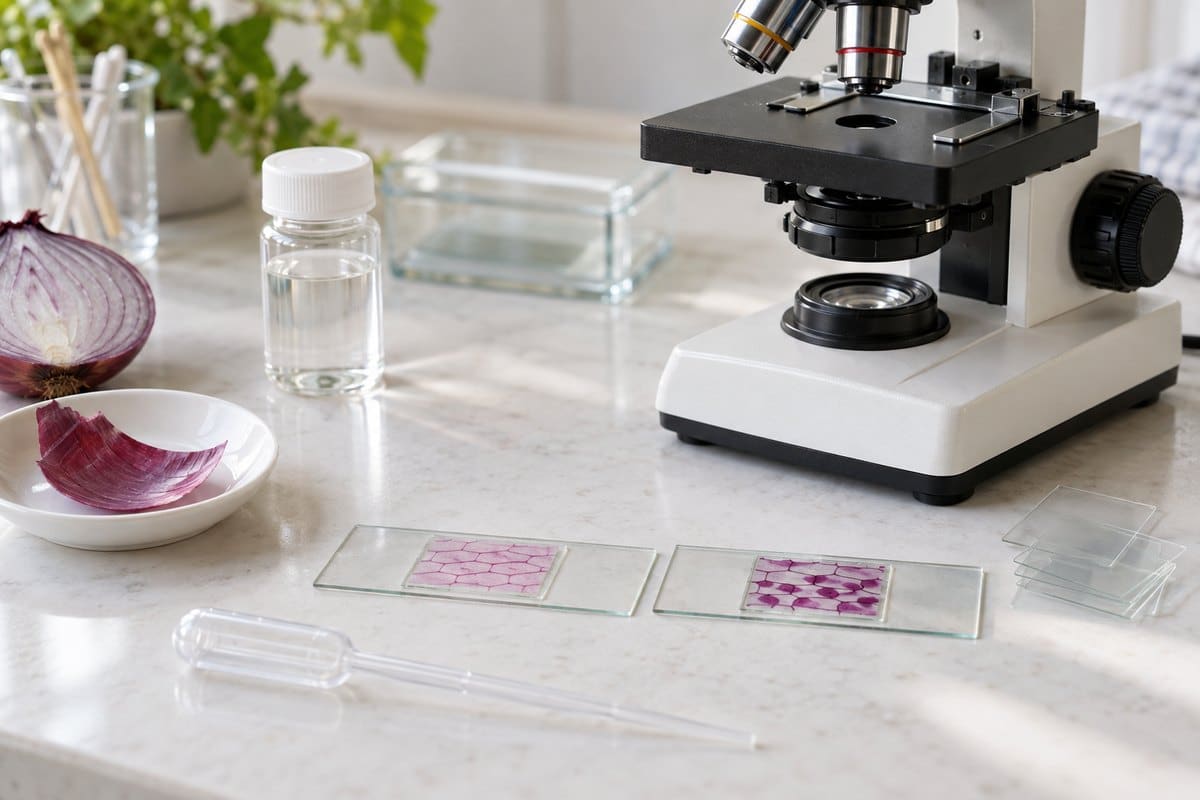
Since the bilayer itself sits below what a light microscope can resolve, the way most students actually “see” the cell membrane is by watching what it does under osmotic stress. This is where the abstract diagram turns into something you can observe on a slide in real time. In plain water, an onion cell’s purple-tinged cytoplasm fills the rigid, rectangular cell wall right up to the edge. The membrane and the wall look like a single line, with nothing to distinguish them. That changes the moment you draw a drop of salt water under the coverslip and watch a salt-soaked onion cell plasmolyze in real time. Over the next minute or two, the colored contents shrink inward and ball up, pulling away from the stiff wall outline and leaving a visible clear gap. That retreating edge is the membrane, and the process is called plasmolysis. It looks like the cell’s insides are deflating away from their own frame.
The same physics plays out in animal cells, just without a wall to hold the shape. Drop red blood cells into pure water and they swell into round balls, then vanish into faint “ghost” rings as they burst — hemolysis. Put them in salty water instead and they shrink and go spiky, edges crinkled like a cog — crenation. You can watch this directly by comparing red blood cells in a hypotonic solution against a normal, isotonic sample side by side. The tonicity behind both reactions comes down to three states. Isotonic means equal solute inside and out, so there’s no net water movement. Hypotonic means more water outside the cell, so water rushes in and the cell swells (or, in a plant cell, becomes turgid, held in check by the wall). Hypertonic means more solute outside, so water leaves and the cell shrivels or, in a plant cell, plasmolyzes.
Three mistakes trip up most beginners here. First, expecting to see the phospholipid bilayer itself — you can’t, since it’s roughly 7 nm thick against a ~200 nm resolution limit. Look for what the membrane does instead of the line itself. Second, confusing the thick, bold cell wall outline with the membrane in plant cells. The wall is the bold line you notice first, but the membrane only becomes visible once salt water pulls it inward, away from the wall. Third, flooding the slide with too much salt at once, which collapses the cell almost instantly and skips right past the gradual pull-away that makes plasmolysis worth watching. A milder solution observed over two to three minutes shows the process properly. Work at 400x, draw the salt solution across the slide with a paper towel wicking from the far side of the coverslip, and don’t look away. The pull-away typically starts within 30 to 60 seconds. If you want to build the slide yourself first, make a wet mount slide and practice the technique on plain water before introducing salt. You’ll notice the same physics outside the lab, too — it’s why lettuce crisps back up in ice water and wilts in salad dressing. It’s also the same mechanism behind a paramecium’s contractile vacuole bailing out water to keep from bursting in its freshwater pond.
Frequently Asked Questions
Is the cell membrane the same thing as the cell wall?
No. Every cell — plant, animal, fungal, bacterial — has a cell membrane, but only plants, fungi, bacteria, and some protists also have a cell wall. The wall sits outside the membrane as an extra rigid layer. The membrane is the universal boundary; the wall is optional extra reinforcement.
Do all living cells have a cell membrane?
Yes. Both prokaryotic cells (like bacteria) and eukaryotic cells (like plant, animal, and fungal cells) are enclosed by a cell membrane. It’s one of the defining features of what counts as a cell in the first place.
Can you see the cell membrane under a school microscope?
Not directly. At roughly 7–8 nanometers thick, the bilayer is far below the resolving power of a standard light microscope. What you actually see are its effects — plasmolysis in a salted onion cell, or swelling and crenation in a red blood cell.
Is “cell membrane” the same as “plasma membrane”?
Yes, the two terms are interchangeable and refer to the same structure. “Cell membrane” is the more commonly used term in US schools; “plasma membrane” shows up more often in college-level and research literature.
How thick is the cell membrane?
The cell membrane is typically cited at about 7–8 nanometers thick. Estimates in the literature range from roughly 5 to 10 nanometers depending on the cell type and measurement method.
What happens if the cell membrane is damaged?
Small tears can often reseal on their own because the bilayer is fluid, not rigid — nearby phospholipids drift in to patch the gap. Larger or sustained damage disrupts the membrane’s ability to control what enters and leaves, which can trigger swelling, leakage of cell contents, and cell death.
Conclusion
The cell membrane looks simple in a textbook diagram — a bilayer, a few proteins, some cholesterol — but every piece of that structure earns its place by doing a specific job. That ranges from the phospholipids’ self-arranging chemistry to the sodium–potassium pump spending ATP to keep a nerve cell’s electrical gradient intact. Once you connect the fluid mosaic model to something you can actually watch happen, like an onion cell plasmolyzing in salt water, the abstract diagram stops being something to memorize. It starts being something you understand.
Have you watched plasmolysis happen on your own slide, or compared hemolysis and crenation side by side under your scope? Tell us what you saw in the comments below — and if something in your sample didn’t match what you expected, that’s usually the most interesting part to dig into.