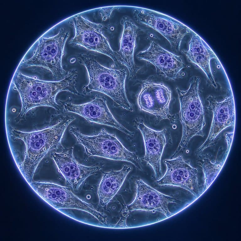
HeLa cells under a microscope look like flat, irregular, glassy patches clinging to the floor of a lab culture flask — not the neat stained specimen most people picture. HeLa is the first immortal human cell line, established in 1951 from the cervical cancer of Henrietta Lacks, and it is still dividing today in laboratories around the world. Immortality here comes down to a hijacked cell-division clock. Telomerase keeps rebuilding the cell’s telomeres, while a virus-driven shutdown of two tumor-suppressor proteins removes the brakes that would normally stop it.
What Are HeLa Cells?
HeLa cells are an immortal human cell line derived from a cervical tumor biopsy taken from Henrietta Lacks in 1951 at Johns Hopkins Hospital. Researcher George Otto Gey cultured the sample, and unlike every human cell line attempted before it, the cells kept dividing indefinitely instead of dying out after a few generations. The tumor was originally recorded as an epidermoid (squamous) carcinoma; later re-examination reclassified it as a cervical adenocarcinoma, and modern sources use that corrected diagnosis. The cancer itself was driven by human papillomavirus type 18 (HPV-18), whose DNA is permanently integrated into the HeLa genome. The NLM MeSH record identifies HeLa as the first continuously cultured human malignant cell line.
Where the Name Comes From
“HeLa” is built from the first two letters of Henrietta Lacks’s first and last names. It was a naming convention Gey’s lab used for cell samples generally, not a tribute created after the fact. For decades the person behind the initials was hidden or misreported in scientific writing, which is part of why getting her story right now matters.
| Trait | HeLa Cells | Normal Human Cell |
|---|---|---|
| Division limit | None (immortal) | ~40–60 divisions (Hayflick limit) |
| Chromosome count | ~76–80 (hypertriploid, aneuploid) | 46 (diploid) |
| Contact inhibition | Largely lost | Intact |
| Doubling time | ~20–24 hours | Varies; slows with senescence |
| Typical spread size | ~20–40 µm | Varies by cell type |
Why HeLa Cells Are Immortal
Normal human cells stop dividing after a set number of divisions, a ceiling known as the Hayflick limit. That happens because their telomeres — protective caps on the ends of chromosomes — shorten a little with every division cycle until the cell enters replicative senescence and stops. HeLa cells bypass that ceiling through two combined mechanisms:
- Telomerase reactivation. HeLa cells express high levels of telomerase, the enzyme that rebuilds telomeres after each division. With telomeres maintained instead of eroding, the countdown to senescence never runs out.
- Tumor-suppressor shutdown. HPV-18 produces the oncoproteins E6 and E7, which inactivate the p53 and retinoblastoma (Rb) proteins — the cell’s own checkpoints for stopping damaged or abnormal division. With those brakes disabled, the cell keeps dividing even when it shouldn’t.
Telomerase is the engine; the E6/E7 disruption of p53 and Rb is the removed brake. Both have to be in place — telomerase alone does not explain HeLa’s behavior. This two-part mechanism is why the cell line has stayed useful in cancer research for over seventy years.
What HeLa Cells Look Like Under a Microscope
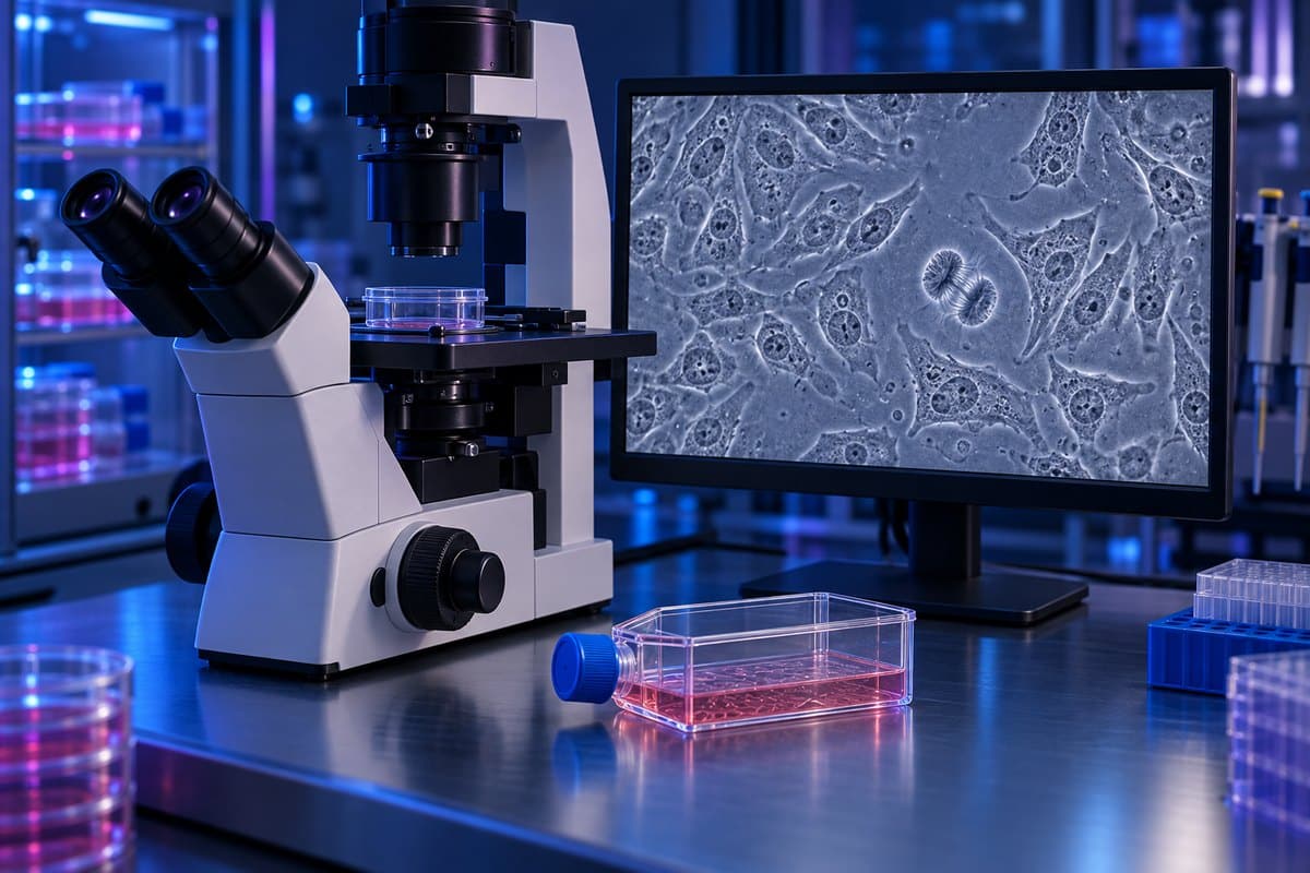
HeLa cells are never viewed on a home microscope slide the way human cheek cells under a microscope or onion cells under a microscope can be. They exist only as a living lab culture in a sealed flask. Researchers examine them with a phase-contrast microscope from below (an inverted setup), since the flask itself sits between the lens and the cells.
In an Unstained Live Culture (Phase Contrast)
At low magnification, a healthy HeLa flask looks like a field of flat, gray, faintly translucent patches stuck to the plastic floor — something close to crazy paving, or a tray of overlapping fried eggs. Each patch is a single cell that has spread out and gone epithelial-like and polygonal, its edges ragged where it presses against its neighbors. Inside each one sits a denser gray oval, the nucleus, usually holding one to four small dark dots — the nucleoli.

Scattered among the flat cells are bright, perfectly round balls, some floating free and some loosely tethered to the surface. A beginner’s first instinct is to assume these round cells are the normal ones and the flat ones are somehow wrong — it is the opposite. The rounded shape means a cell has either pulled itself up to divide (mitosis makes cells detach and go spherical) or has died and lost its grip on the flask. A culture full of round floaters and thin on flat, spread cells is a stressed or overgrown one, not a thriving one.
Left to grow, the flat cells eventually pack edge to edge into a seamless cobblestone sheet with no gaps between them — confluence. Because HeLa cells have largely lost contact inhibition, pushing past that point doesn’t stop growth the way it would in a normal cell line. The sheet starts heaping into disorganized 3D clumps instead, and researchers split (“passage”) the culture before that happens to keep it healthy.
In Stained and Fluorescence Images
The colorful HeLa images most people have actually seen — a glowing blue nucleus over a web of green or red cytoskeleton — come from fixed, stained, or fluorescently labeled cells. They are not the living culture. A common combination stains the nucleus deep blue with DAPI while a fluorescent tag lights up a specific structure, like actin filaments or microtubules, in a contrasting color. This kind of imaging requires fluorescence and confocal microscopy and kills the cells in the process. That means it captures a single frozen moment rather than the ongoing, ghostly gray movement you’d see live. HeLa’s reliability and flat, spread-out shape have also made it the standard test subject for newer techniques like super-resolution microscopy techniques. Its well-defined structures are ideal for checking image resolution.

Who Was Henrietta Lacks?
Henrietta Lacks was a Black woman, wife, and mother of five from Baltimore. She sought treatment for abnormal vaginal bleeding at Johns Hopkins Hospital — one of the few hospitals in the region that treated Black patients at the time. Doctors diagnosed her with cervical cancer, and during a biopsy in 1951, a sample of her tumor tissue was sent to George Gey’s lab without her knowledge or consent. That was a common and legal practice for the era, but one that would not meet today’s research ethics standards. She died from the cancer on October 4, 1951, at age 31. NIH documents the origin of HeLa cells and the NIH–Lacks Family Agreement.
Neither Henrietta Lacks nor her family were told that her cells had been taken, or that they were being grown, sold, and shipped to labs worldwide. For decades the family saw no compensation while HeLa cells generated enormous scientific and commercial value. Her identity was also mishandled in public: her cells were misattributed to other names in early scientific writing, stripping her of recognition for years. Journalist Rebecca Skloot’s 2010 book The Immortal Life of Henrietta Lacks brought her story to wide public attention. In 2013, the Lacks family reached an agreement with the NIH giving them a say in how HeLa genomic data is accessed. In 2023, the family reached a settlement with Thermo Fisher Scientific over the commercial use of her cells. According to Johns Hopkins Medicine’s account of her legacy, the institution now formally recognizes the ethical debt owed to her and her family.
Why HeLa Cells Matter
HeLa cells were the first cells mass-cultured for vaccine research, making Jonas Salk’s polio vaccine trials possible in the 1950s. Since then they’ve been used to study cancer biology, virology, gene mapping, toxicology, and the effects of spaceflight on human cells. More recently, researchers have used them to study how the SARS-CoV-2 spike protein interacts with the ACE2 receptor. According to the National Cancer Institute, HPV-driven cancers like the one that produced HeLa remain a major focus of ongoing research. That is part of why this specific cell line has stayed relevant for so long.
That same aggressive growth has a downside: HeLa cells are notorious lab contaminants. Because they divide so fast and tolerate a wide range of conditions, stray HeLa cells have invaded and silently overtaken hundreds of other “unique” cell lines in labs worldwide over the decades. That problem is serious enough that researchers now routinely genetically verify a culture’s identity before trusting results from it.
Frequently Asked Questions
Are HeLa cells cancer cells?
Yes. They originated from a cervical adenocarcinoma, a form of cervical cancer, which is why they divide uncontrollably and ignore the normal signals that would stop a healthy cell. This article covers the science and history of HeLa cells for educational purposes and isn’t medical advice about cancer or HPV.
How many chromosomes do HeLa cells have?
HeLa cells are hypertriploid and aneuploid, typically carrying around 76 to 80 chromosomes, compared with the normal human count of 46. The exact number varies somewhat between HeLa sub-lines because the genome is unstable.
Are HeLa cells still used today?
Yes, extensively. They remain a standard tool in cancer research, virology, and cell biology, and have contributed to recent work on COVID-19 and the ACE2 receptor.
Did Henrietta Lacks or her family consent to the use of her cells?
No. The cells were taken and cultured without her knowledge or consent, and her family was not informed for decades. Agreements in 2013 (with the NIH) and 2023 (with Thermo Fisher Scientific) later addressed data access and commercial use.
How big are HeLa cells?
A spread, adherent HeLa cell typically measures around 20 to 40 micrometers (µm) across. Rounded cells that are dividing or detaching appear noticeably smaller and brighter under phase contrast.
Conclusion
HeLa cells are a case where the microscope image and the human story are inseparable. The flat, crowded, cobblestone sheet you’d see under phase contrast exists only because telomerase and a disabled p53/Rb checkpoint let a single cancer cell from Henrietta Lacks keep dividing since 1951. Understanding what these cells look like in culture, and what they don’t look like since they’re never a home slide specimen, makes the underlying biology easier to grasp. It also makes the ethics behind their story easier to hold onto.
Have you come across HeLa cells in a biology class, textbook, or Rebecca Skloot’s book? Share what stood out to you about their story in the comments below.