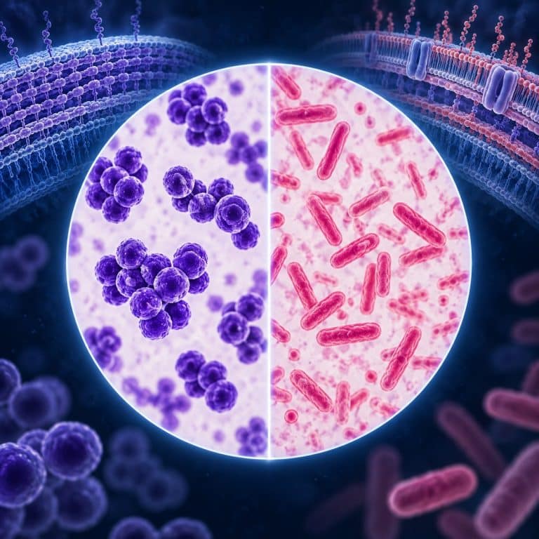
Gram-positive and gram-negative bacteria differ mainly in their cell wall structure, and that single structural difference explains almost everything else that separates them. Gram-positive cells wrap a thick peptidoglycan layer around a single membrane, trapping the purple crystal violet stain during a Gram stain. Gram-negative cells carry a thin peptidoglycan layer sandwiched between two membranes, so the same stain washes back out and the cells pick up a pink counterstain instead.
The Core Difference: Cell Wall Structure
The cell envelope is where gram-positive and gram-negative bacteria part ways. Gram-positive bacteria are monoderms — a single plasma membrane wrapped in one thick peptidoglycan wall. Gram-negative bacteria are diderms — a thin peptidoglycan wall sandwiched between an inner plasma membrane and a second, outer membrane. That extra membrane, not the peptidoglycan alone, is what drives most of the practical differences covered below, from staining behavior to antibiotic resistance patterns in gram-negative infections. OpenStax diagrams the different cell-envelope structures of Gram-positive and Gram-negative bacteria.
Gram-Positive Cell Wall
A gram-positive cell wall is built from a peptidoglycan (murein) layer roughly 20 to 80 nanometers thick. That’s the same thick layer that Carleton College’s microbial life resource describes as what retains the crystal violet stain during Gram staining. Woven through that peptidoglycan are wall teichoic acids, anchored directly to the peptidoglycan mesh, and lipoteichoic acids, anchored to the plasma membrane underneath. Both are unique to gram-positive cells and help the wall hold its shape and regulate ion movement. There is no outer membrane at all — the thick peptidoglycan layer is the outermost structure a gram stain reagent has to get through.
Gram-Negative Cell Wall
A gram-negative cell wall is almost the opposite arrangement: a thin peptidoglycan layer, only about 2 to 7 nanometers thick, sitting inside a fluid-filled periplasmic space. Wrapped around that periplasm is an outer membrane, and the outer leaflet of that membrane is built from lipopolysaccharide (LPS) rather than ordinary phospholipid. Studded through the outer membrane are porins — barrel-shaped channel proteins that control which molecules, including many antibiotics, can pass through. No teichoic acids are present. This extra membrane is the main reason gram-negative bacteria behave so differently from gram-positive bacteria in a lab or a clinic.
Gram Positive vs Gram Negative: Comparison Table
Here is the full picture side by side. If you only memorize one thing from this article, memorize this table.
| Property | Gram-Positive | Gram-Negative |
|---|---|---|
| Peptidoglycan layer | Thick (~20–80 nm) | Thin (~2–7 nm) |
| Outer membrane | Absent | Present (with LPS) |
| Membrane count | Monoderm (1) | Diderm (2) |
| Teichoic acids | Present | Absent |
| Periplasmic space | Minimal or none | Prominent |
| Gram stain color | Purple/violet | Pink/red |
| Endotoxin (LPS/lipid A) | No | Yes |
| Example genera | Staphylococcus, Streptococcus, Bacillus | E. coli, Salmonella, Pseudomonas |
| General antibiotic response | Often penicillin/vancomycin-susceptible | Often more resistant (outer-membrane barrier) |
Why One Stains Purple and the Other Pink
The Gram stain result is not arbitrary — it is a direct readout of the cell wall structure above, and the American Society for Microbiology’s Gram stain protocol lays out the sequence clearly: Its staining guide also explains the crystal-violet, iodine, decolorizer and safranin steps of the Gram stain.
- Crystal violet is applied first and stains every cell, gram-positive or gram-negative, purple.
- Gram’s iodine acts as a mordant, binding to the crystal violet to form a large crystal-violet–iodine (CV-I) complex trapped inside the cell.
- Decolorizer (alcohol or acetone) is the deciding step. In gram-positive cells it dehydrates and shrinks the thick peptidoglycan mesh, tightening the pores and trapping the CV-I complex inside — the cell stays purple. In gram-negative cells it dissolves the lipid-rich outer membrane instead, and the CV-I complex simply washes out through the thin, now-exposed peptidoglycan.
- Safranin, a pink counterstain, is applied last. It has no effect on gram-positive cells, which are already saturated purple, but it stains the now-colorless gram-negative cells pink to red.
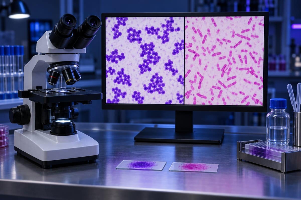
The result: gram-positive bacteria read purple or violet, gram-negative bacteria read pink or red. Under oil immersion at 1000x, the color difference is genuinely stark once the timing is right. Staphylococcus shows up as deep purple grape-like clusters, Streptococcus as purple bead-chains, while gram-negative rods and cocci sit pale pink against the field and are easy to lose if you over-decolorize.

The single most common mistake beginners make is leaving the decolorizer on too long. A few seconds too many strips the crystal violet out of gram-positive cells as well, and a genuinely gram-positive organism reads back as a false pink. The fix is to decolorize only until the runoff coming off the slide turns clear, then rinse immediately. The whole result can live or die in about five seconds of alcohol. A second common error is smearing the sample too thick. Dye pools unevenly, so the center of the smear reads purple while the thinner edges of the very same organism read pink. A thin, even smear read at the edges gives the truest result. Aging cultures cause a third failure mode. Bacteria more than about 24 hours old can lose wall integrity and stain gram-variable, patchy purple and pink on the same slide, which is why fresh cultures matter for a reliable read.
Examples of Each Type
Both groups span common genera you have likely already encountered, and what bacteria look like under a microscope in general is a good primer before comparing species side by side.
| Genus | Gram Status | Shape | Where You Might See It |
|---|---|---|---|
| Staphylococcus | Positive | Coccus (clusters) | Skin flora; S. aureus |
| Streptococcus | Positive | Coccus (chains) | Throat, skin infections |
| Lactobacillus | Positive | Rod | You can see the Lactobacillus in yogurt at home |
| Escherichia | Negative | Rod | E. coli, a classic gram-negative rod |
| Salmonella | Negative | Rod | See Salmonella under the microscope |
| Neisseria | Negative | Coccus | Respiratory tract |
Notice that shape does not track with Gram status. A common student mix-up is assuming “positive means round, negative means rod-shaped” — but Bacillus is a gram-positive rod and Neisseria is a gram-negative coccus. Shape and Gram classification are two completely independent properties of a cell.

Why the Difference Matters (Antibiotics & Infection)
This section is educational content, not medical advice. Antibiotic choice is always a clinical decision made by a qualified provider, and the patterns below are general tendencies, not treatment rules.
The outer membrane is the reason gram-negative and gram-positive infections are often managed differently. Gram-positive bacteria expose their thick peptidoglycan wall directly to the environment. That’s why beta-lactam antibiotics like penicillin and glycopeptides like vancomycin, drugs that target peptidoglycan synthesis, tend to work well against them. Gram-negative bacteria hide that same peptidoglycan behind an outer membrane whose porins restrict what can get through, so many antibiotics that work well on gram-positive cells struggle to reach their target at all. It’s a mechanism CDC’s overview of gram-negative bacteria identifies as a major driver of drug-resistant infections.
The outer membrane’s LPS layer adds a second clinical dimension. LPS is built from lipid A, a core oligosaccharide, and an O-antigen chain. The lipid A portion functions as an endotoxin — a toxic compound released when a gram-negative cell lyses. Research on endotoxin’s role in sepsis shows that large amounts of released LPS can trigger an exaggerated inflammatory response, fever, and, in severe cases, organ injury from septic shock. Gram-positive cells have no LPS and do not carry this particular risk.
None of this makes one group universally “worse.” Resistance patterns like MRSA (methicillin-resistant Staphylococcus aureus), VRE (vancomycin-resistant Enterococcus), and ESBL/CRE (extended-spectrum beta-lactamase and carbapenem-resistant Enterobacteriaceae) gram-negative strains regularly break the general pattern. Both groups contain species that are harmless and species that are dangerous pathogens. The gram-positive/gram-negative split guides antibiotic selection; it does not dictate it.
Exceptions: Bacteria That Don’t Fit
Not every bacterium sorts cleanly into one column. Gram-variable species stain inconsistently — sometimes from genuine intermediate wall structure, sometimes from the aging-culture problem described above. Acid-fast bacteria such as Mycobacterium (the genus behind tuberculosis) have a waxy, mycolic-acid-rich wall that resists standard Gram staining altogether and requires a separate acid-fast stain. Wall-less bacteria like Mycoplasma have no peptidoglycan to react with crystal violet in the first place, so a Gram stain simply cannot classify them. These exceptions are a useful reminder that the Gram stain is a chemistry test on a specific structure, not a universal bacterial fingerprint.
Frequently Asked Questions
Is E. coli gram-positive or gram-negative?
Gram-negative. E. coli has the thin peptidoglycan wall and outer LPS membrane typical of the group, and it stains pink under a Gram stain.
Is Staphylococcus gram-positive or gram-negative?
Gram-positive. Staphylococcus has a thick peptidoglycan wall with no outer membrane and stains purple, typically forming the grape-like clusters the genus is known for.
Can a bacterium be neither gram-positive nor gram-negative?
Yes. Gram-variable species stain inconsistently. Acid-fast bacteria like Mycobacterium resist the stain because of their waxy wall, and wall-less bacteria like Mycoplasma have no peptidoglycan for the stain to react with at all.
What does the Gram stain actually test?
It tests how much peptidoglycan a cell wall has, and whether an outer membrane is present. Thick peptidoglycan without an outer membrane traps the stain and reads purple; thin peptidoglycan with an outer membrane loses the stain and reads pink.
How long does a Gram stain take from start to finish?
A full slide, from heat-fixing the smear through crystal violet, iodine, decolorizer, and safranin, typically takes five to ten minutes. The decolorizer step itself is the only part where timing is critical, often just a few seconds.
Can a gram-positive bacterium turn into a gram-negative one, or vice versa?
No. Cell wall type is a stable structural feature of a species and does not switch. What looks like a change is almost always a staining or technique problem — an old culture, too thick a smear, or over-decolorizing. That produces a misleading result rather than a real structural shift.
What magnification do I need to see Gram stain results clearly?
Oil immersion at 1000x is standard. At lower power you can make out clumps of color, but you cannot resolve individual cells well enough to judge purple versus pink with confidence. That’s especially true on gram-variable or lightly stained slides.
Conclusion
Every difference in this article traces back to one structural fact. Gram-positive bacteria wrap a thick peptidoglycan wall around a single membrane, while gram-negative bacteria wrap a thin wall and an extra outer membrane around theirs. That one distinction decides the stain color, shapes which antibiotics work best, and explains why gram-negative infections carry the added risk of endotoxin release. Keep the comparison table above handy and the rest follows logically from it.
Have you run a Gram stain yourself, in a classroom or at home with a prepared slide? Share what you saw, or what tripped you up on your first try, in the comments below.