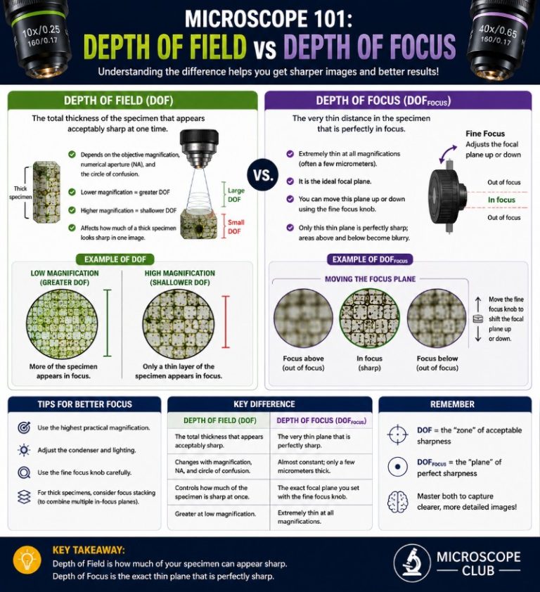
Depth of field in a microscope is the axial distance — measured along the optical axis — over which a specimen remains in acceptable focus without repositioning the objective or stage. At a 100x oil-immersion objective it can be as thin as 0.2 µm, which means a single bacterium can slip in and out of focus while you adjust the coarse knob. Understanding what controls that razor-thin band, and how to work around it, is one of the most practical things you can learn at the bench.
What is depth of field in a microscope?

Depth of field is the range along the optical axis within which the specimen produces an acceptably sharp image. It is the axial (longitudinal) resolving power of the objective lens — different from lateral resolution, which is the ability to distinguish two side-by-side points. In practice, depth of field tells you how much you can move the stage up or down before the image blurs.
Because depth of field is measured in the object space (at the specimen), it is strongly influenced by the objective’s numerical aperture (NA). Typical values run from ~55 µm at 4x (NA 0.10) down to well under 1 µm at 100x oil immersion (NA 1.25–1.4). At those figures, auto-focus systems or careful motorized control become nearly essential — the hand is rarely steady enough at high magnification to hold focus manually for any useful length of time.
What is depth of focus?
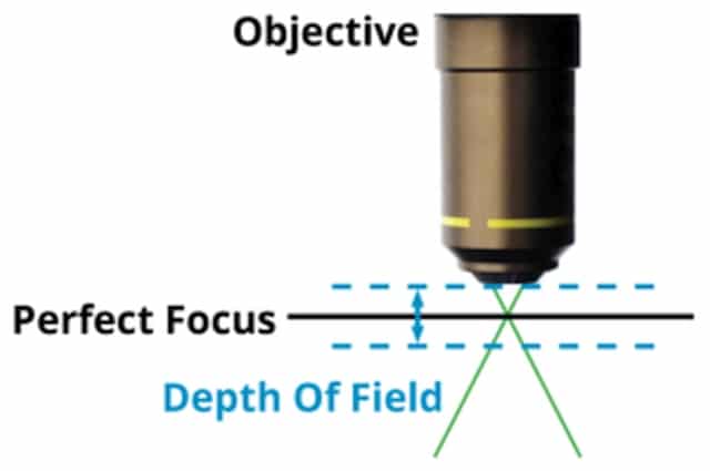
Depth of focus is the image-space counterpart of depth of field. While depth of field describes how far the specimen can move and still appear sharp, depth of focus describes how far the detector — camera sensor or eyepiece plane — can shift along the optical axis before the image degrades. The two are related but distinct: a thick specimen challenges depth of field, while a slightly misaligned camera back challenges depth of focus.
Depth of focus increases with magnification — a high-magnification objective gives a deep image-side focus range even as the object-side depth of field narrows dramatically. It also scales with the square of the objective’s NA as seen from the image side. For digital cameras, pixel pitch directly sets the minimum resolvable detector distance e — a camera with larger pixels is more forgiving of small focus-plane offsets. Sensors with 3–5 µm pixel pitch are typical in research cameras, and their influence shows up in the combined depth of field formula in the second term.

What factors affect depth of field?
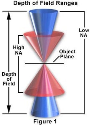
Four variables drive depth of field. Changing any one of them shifts the in-focus band — sometimes dramatically. The key one to internalize first is numerical aperture, because it appears squared in the denominator of the formula.
Numerical aperture
Numerical aperture (NA) is defined as n·sin(θ), where n is the refractive index of the medium between specimen and objective and θ is half the angular cone of light the objective accepts. Depth of field is inversely proportional to NA² — double the NA and the depth of field shrinks to roughly one quarter. This also means that raising NA to improve resolution always costs depth of field. That trade-off is built into the physics and cannot be bypassed optically (though focus stacking and confocal techniques work around it after the fact).

Resolution and NA — the core trade-off
A common misconception in older microscopy texts is that lower NA gives both better resolution and deeper depth of field. This is wrong. Higher NA yields finer (better) lateral resolution — the resolvable distance between two points shrinks as NA rises — while depth of field simultaneously narrows. The relationship is an unavoidable trade-off: high NA → fine resolution, shallow depth of field; low NA → poor resolution, deep depth of field — confirmed across major manufacturers (Evident/Olympus NA primer; Basler).
Contrast and specimen thickness
High-contrast specimens with sharp boundaries look sharp across a slightly wider axial range because the eye and camera pick up any residual edge detail more readily. Low-contrast specimens — unstained cells in phase contrast, for example — can appear to go soft sooner. Specimen thickness is the direct practical consequence: a thin smear (blood film, bacteria) stays fully in the depth of field band even at 100x; a thick section (50 µm plant tissue, whole mount) will require focusing through multiple planes, since no single focus position renders the entire specimen sharp simultaneously.
Working distance
Working distance is the free space between the front element of the objective and the cover slip when the specimen is in focus. Shorter working distance goes hand in hand with higher NA (the objective must get closer to collect a wide light cone), which in turn reduces depth of field. A 100x oil objective typically has a working distance of 0.1–0.17 mm; a 4x objective may have several millimeters. The shorter the working distance, the more precise the z-axis control needs to be.
Magnification
Increasing magnification reduces depth of field, partly because higher magnification objectives generally carry higher NA, and partly because the geometric term in the combined formula is divided by magnification. At low magnification (4x, 10x), thick and irregularly shaped objects are generally manageable — most of the specimen sits within the generous depth of field. At 40x and above, a single pollen grain (~20 µm tall) extends beyond the in-focus band, and you will need to rack through focus to examine it fully.
Immersion media and refractive index
The refractive index n of the medium between specimen and objective enters both the wave-optical and geometric terms of the depth of field formula. Air has n = 1.00 (the theoretical baseline). Water immersion objectives use n ≈ 1.33, which raises achievable NA and slightly deepens the wave-optical depth of field term compared to air at the same NA. Immersion oil (typically n ≈ 1.515) raises NA further — a 100x oil objective can reach NA 1.25–1.4, far beyond what is possible in air (theoretical dry maximum ≈ 1.0), as detailed in ZEISS’s oil immersion lens design guide. The practical payoff is finer resolution at the cost of a shallower in-focus band.
How to calculate microscope depth of field
Two formulas are in common use, matching two different physics regimes.
Diffraction-limited (wave-optical) depth of field
At any magnification, the wave-optical contribution to depth of field is:
dwave = (λ · n) / NA²
where λ is the wavelength of the illuminating light (enter in µm), n is the refractive index of the medium, and NA is the numerical aperture. This is the standard diffraction-limited wave-optical formula.
Combined (total) depth of field — for high magnification
At high magnification the detector’s finite resolution also matters. The complete formula adds a geometric term:
dtot = (λ · n / NA²) + (n / (M · NA)) · e
where M is the lateral magnification of the objective and e is the smallest resolvable distance of the detector in the image plane — approximately 1.5 µm for the human eye at the eyepiece, or the camera’s pixel pitch for digital cameras (commonly 3–6 µm for research-grade sensors).
Worked example — 40x dry objective
Let’s walk through both terms using a common 40x plan-apochromat:
- Objective: 40x dry, NA = 0.65, so n = 1.00 (air)
- Illumination: green light, λ = 0.55 µm
- Detector: visual observation, e = 1.5 µm; M = 40
Wave-optical term: dwave = (0.55 × 1.00) / 0.65² = 0.55 / 0.4225 ≈ 1.30 µm
Geometric term: (1.00 / (40 × 0.65)) × 1.5 = (1.00 / 26) × 1.5 ≈ 0.06 µm
Total: dtot ≈ 1.36 µm — consistent with the MicroscopyU reference table value of ~1.0 µm for NA 0.65 (minor variance reflects different e assumptions). At this scale, moving the stage by as little as 1–2 µm will push fine specimen detail out of focus.
If you swap to a camera with 5 µm pixels, the geometric term rises to (1.00 / 26) × 5 ≈ 0.19 µm — still dominated by the wave term at this NA, but the balance shifts more at lower magnification where NA is smaller and M is lower.
Depth of field reference table
The table below shows typical depth of field values calculated for green light (λ = 0.55 µm) using common objective specifications. Use it as a quick-reference for your own setup, adapted from the Nikon MicroscopyU depth of field reference.
| Magnification | Typical NA | Depth of Field (µm) | Depth of Focus (mm) |
|---|---|---|---|
| 4x | 0.10 | ~55.5 | 0.13 |
| 10x | 0.25 | ~8.5 | 0.80 |
| 20x | 0.40 | ~5.8 | — |
| 40x | 0.65 | ~1.0 | 12.8 |
| 100x (dry) | 0.95 | ~0.19 | 80.0 |
Standard objective NA values: 10x ≈ 0.25, 20x ≈ 0.50–0.75, 40x ≈ 0.65, 100x oil immersion ≈ 1.25–1.4 (high-end designs reach 1.45; theoretical maximum with standard oil is approximately 1.51).
Stereo vs compound microscope: a depth of field contrast
Stereo microscopes and compound microscopes sit at opposite ends of the depth-of-field spectrum — a contrast covered in detail in the Leica Science Lab depth of field guide. Understanding why explains a lot about which tool to reach for.
A stereo microscope (dissecting microscope) uses low-NA optics — typically NA 0.05–0.15 — designed to keep large, three-dimensional objects in simultaneous focus. At 10–45x, depth of field runs from roughly 1–3 mm, which is why you can inspect a circuit board or dissect a flower and see the whole specimen sharp at once. A stereo microscope is optimized for object-space depth, trading away fine lateral resolution to achieve it.
A compound light microscope at 40x or 100x has depth of field measured in single-digit microns. That shallowness is what makes it possible to resolve subcellular structures — the narrow depth of field and high NA come as a package. The trade-off is that thick specimens require systematic focus-through to build a mental three-dimensional picture.
| Feature | Stereo microscope | Compound microscope (high mag) |
|---|---|---|
| Typical NA | 0.05–0.15 | 0.65–1.4 |
| Depth of field | 1–3 mm | 0.2–1.5 µm |
| Best for | Large, 3D objects; dissection; assembly | Thin sections; cells; subcellular detail |
How to increase depth of field: focus stacking (z-stacking)
Focus stacking — also called z-stacking — is the standard technique microscopists use to beat the physical limits of shallow depth of field. The approach is post-acquisition: you capture a series of images at evenly spaced focus positions along the z-axis, stepping through the specimen in increments smaller than the objective’s depth of field. Software then merges the sharpest pixels from each frame into a single composite image that shows the full three-dimensional specimen in sharp focus, even when no single focus position achieves that optically.
The step interval matters — too large and you leave z-gaps; too small and you burn acquisition time and increase bleaching risk in fluorescence. A common starting rule: set the z-step to roughly half the depth of field. For a 40x/0.65 objective (~1 µm DoF), 0.5 µm steps are appropriate. Software options include Helicon Focus, Zerene Stacker, Fiji/ImageJ (Extended Depth of Focus plugin), and built-in acquisition tools in platforms like Leica LAS X and Zeiss ZEN.
Confocal microscopy and depth of field
Confocal microscopy takes the depth-of-field problem and inverts it into a feature. A confocal system places a pinhole aperture at the back focal plane, physically blocking out-of-focus light before it reaches the detector. This reduces the effective depth of field to ~0.5 µm or less — far narrower than a conventional widefield objective at the same NA — allowing optical sectioning: collecting a stack of perfectly defined planes through a thick specimen without the haze from planes above and below.
The result is that confocal is the standard tool for 3D fluorescence imaging of specimens too thick for widefield to resolve cleanly. Because each optical section is clean, the z-stack is much easier to process into a 3D reconstruction. Confocal microscopes trade some signal — the pinhole rejects most photons — for dramatically improved axial resolution. It is the depth-of-field/image-quality trade-off that runs through all of microscopy, carried to its logical conclusion.
Frequently asked questions
-
What is the difference between depth of field and depth of focus in a microscope?
Depth of field is an object-space measurement — how far the specimen can move along the optical axis and remain in focus. Depth of focus is an image-space measurement — how far the camera or eyepiece plane can shift before the recorded image degrades. Higher
magnification deepens depth of focus even as it narrows depth of field.
-
Does higher magnification increase or decrease depth of field?
Higher magnification decreases depth of field. Both the wave-optical term (through higher NA) and the geometric term (through the 1/M factor) shrink the in-focus band as magnification rises.
-
How does numerical aperture affect depth of field?
Depth of field is inversely proportional to NA². Doubling the NA reduces depth of field by approximately four times. This is why oil immersion objectives (NA up to 1.4) have depths of field measured in tenths of a micron.
-
Why do stereo microscopes have greater depth of field than compound microscopes?
Stereo microscopes use low-NA optics (NA 0.05–0.15) designed for viewing large 3D objects. Low NA means the light cone is narrow, and the in-focus band is deep — often 1–3 mm. Compound microscopes use high-NA objectives to achieve fine resolution, which inherently compresses the depth of field to the micron range.
-
How do you increase the depth of field of a microscope?
Three approaches: (1) Switch to a lower-magnification, lower-NA objective — the simplest optical fix. (2) Close the condenser aperture diaphragm slightly — this reduces the effective NA at the cost of some resolution and introduces diffraction artifacts if over-closed. (3) Use focus stacking / z-stacking — capture a series of images at stepped focus planes and merge them in software. The third approach is the only one that delivers both fine resolution and extended effective depth of field.
-
What is focus stacking in microscopy?
Focus stacking (z-stacking) is a post-processing technique in which a series of images captured at sequential focus depths are computationally merged, keeping only the sharpest region from each frame. It is the standard workaround for shallow depth of field in macrophotography and high-magnification microscopy, especially for thick specimens or 3D objects.
Conclusion
Depth of field in a microscope is governed primarily by NA² — and because resolution scales with NA in the opposite direction, deeper depth of field always comes at the cost of finer lateral resolution. The practical hierarchy runs from stereo microscopes (millimeter-scale depth of field, modest resolution) through low-power compound objectives (tens of microns) to oil immersion at 100x (tenths of a micron). For thick specimens that exceed any single objective’s depth of field, focus stacking is the standard solution, and for fluorescence work requiring clean optical sections, confocal is the purpose-built answer. Start by reading the reference table above for your objective’s typical depth of field, then use the worked formula to adjust for your specific wavelength and detector — that puts you in the right ballpark before you ever touch the fine focus.