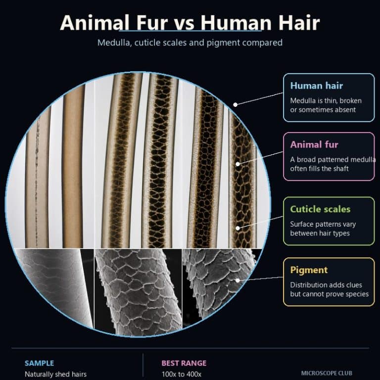
Animal fur vs human hair under a microscope comes down to three structures: the medulla, the cuticle scale pattern, and the way pigment sits inside the shaft. Animal fur usually shows a wide, patterned medulla and crown- or petal-shaped scales, while human hair shows a thin or absent medulla under flat, tile-like scales. Below is a species comparison table for human, cat, dog, rabbit, and mouse hair, plus a slide-prep method that reveals the medulla in minutes.
The Three Features That Separate Fur from Human Hair
Every hair or fur strand is built from the same three layers — cuticle, cortex, and medulla — but the proportions and patterns of those layers differ sharply between species. Forensic examiners and wildlife biologists lean on the same three checkpoints every time: how wide the medulla is and how it’s patterned, what shape the cuticle scales are, and where the pigment granules cluster. Learn to read these three, in order, and you can sort human hair from animal fur with a single glance through the eyepiece.
The Medulla: The Single Most Reliable Clue
The medullary index — the medulla’s diameter divided by the total width of the shaft — is the single most useful number in hair identification. Human hair typically has a medullary index under one-third: the medulla is often fragmentary, broken into disconnected patches, or missing entirely, especially in fine or blonde hair. Animal fur runs the opposite way, with a medullary index above one-half that fills most of the shaft and shows a continuous, species-specific internal pattern. A uniserial ladder (a single stack of rungs) shows up in small rodents and some cats. A multiserial ladder (several parallel rows of cells) is the classic rabbit pattern. Deer and other hoofed animals show a lattice or cellular medulla, while dog hair typically shows an amorphous but wide medulla with no defined internal shape.
Cuticle Scale Pattern: What the Surface Reveals
Cuticle scales form the outermost layer of every hair, and their shape is far more varied on animal fur than on human hair. Human cuticle scales are imbricate — flat, tile-like, and closely overlapping, running in a regular, uniform pattern across the whole shaft. Animal fur breaks that uniformity in three recognizable ways. Coronal scales wrap the shaft like stacked crowns and show up almost exclusively on fine hairs from small mammals such as mice, rats, and bats. Spinous or petal scales protrude in sharp triangular points and are a strong tell for cat and seal fur. Dog and other larger mammals show a looser imbricate/mosaic pattern that can resemble human hair closely enough that scale pattern alone shouldn’t be your only check.
Pigment Distribution and Ovoid Bodies
Pigment (melanin) distribution is the quieter of the three clues, but it’s a useful tie-breaker. In human hair, pigment granules sit evenly through the shaft, or slightly denser toward the outer cuticle edge. In animal fur, pigment tends to concentrate centrally, clustering closer to the medulla, and many species — cow and deer especially — show large, dense, oval pigment clumps called ovoid bodies that essentially never appear in human hair. If you see ovoid bodies on a slide, you’re looking at animal fur, full stop.
Species Comparison Table: Human vs Cat, Dog, Rabbit, and Mouse
The table below lines up the three diagnostic features side by side across five common sources, plus the “standout tell” — the one detail that gives each species away fastest under the scope. If you’ve already worked through a strand of human hair on its own, our full guide to human hair under a microscope covers that baseline in more depth; this table is where you measure everything else against it.
| Feature | Human | Cat | Dog | Rabbit | Mouse |
|---|---|---|---|---|---|
| Medulla (index) | Absent or fragmentary, thin (< ⅓) | Continuous, wide, uniserial ladder | Continuous, wide, amorphous (> ½) | Continuous, multiserial ladder | Continuous, uniserial ladder |
| Cuticle scale pattern | Imbricate (flat tiles) | Spinous/petal | Imbricate/mosaic | Coronal/chevron | Coronal (crown) |
| Pigment | Even, or denser at cuticle | Central, denser at medulla | Central | Central | Central |
| Typical diameter | 50–100 μm | ~30–60 μm | 30–150 μm (guard vs underfur) | ~10–30 μm (underfur) | Fine |
| Standout tell | Air-line, no real medulla | Petal scales + fat medulla | Grainy, wide medulla | Ladder medulla | Coronal crown scales |
Diameter is listed last on purpose — it’s the weakest of the five rows. Human scalp hair (roughly 50–100 μm) overlaps with coarser dog guard hairs at one end and fine cat or rabbit underfur at the other, so “thicker” or “thinner” alone will mislead you more often than it helps.

Guard Hairs vs Underfur: Why Fur Isn’t One Thing
Fur is a two-layer coat, and that’s a structural difference human hair simply doesn’t have. Most mammals grow coarse, longer guard hairs on the outside for weather protection and a soft, fine underfur (or down hair) beneath it for insulation. Human scalp hair has no guard/underfur split at all — every strand grows from broadly the same kind of follicle. That matters for identification: comparing a wisp of rabbit underfur against a strand of human hair and concluding “animal hair is thinner” is a common beginner mistake, because you’re comparing the wrong layer. Always compare a guard hair, not underfur, if you want a fair read against human hair.
How to See the Medulla and Scales Yourself
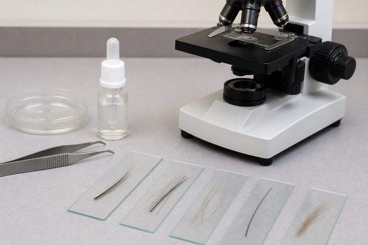
Seeing these features firsthand takes about ten minutes and no special equipment beyond a compound microscope and a bottle of clear nail polish. The most common beginner mistake is viewing a dry hair and mistaking the dark line running down the center for the medulla — that dark line is usually just air trapped inside the shaft, scattering light. Mounting the hair in oil clears that air out and reveals the real internal structure underneath, and the hair almost visibly “switches on” the moment the oil settles in.
- Collect a hair sample. Pull rather than cut if you can — a plucked hair keeps its root, which is a useful extra diagnostic clue (human roots are club-shaped and uniform; animal roots vary by species and growth phase).
- Start with a wet mount slide. Lay the hair flat and straight on the slide — a curled hair drifts in and out of focus and is hard to read.
- Re-mount in immersion oil or a clear mounting medium. This is the step that actually reveals the medulla: the oil fills the air pockets in the shaft so you can see the true internal pattern instead of a trapped-air illusion.
- Make a scale cast for the cuticle. Press the hair into a thin, still-wet layer of clear nail polish, let it dry for two to three minutes, then peel the hair away — the impression left behind shows the scale pattern far more clearly than trying to focus through the hair itself.
- View at 40×–400×. The medulla is visible from 40×–100× once cleared in oil; scale detail is sharpest at 100×–400×. Stop down the condenser or lower the light if the scale edges look blown out.
For the basics of getting a clean, bubble-free slide before you start comparing samples, our guide to preparing microscope slides covers the setup step by step. This video walks through the same human-vs-animal comparison in practice:

How Forensic and Wildlife Scientists Use This
Forensic hair examiners and wildlife biologists both rely on the same three-feature checklist, but for different goals. A wildlife biologist working a poaching or predation case uses the medulla pattern and scale taxonomy to identify which species a tuft of fur came from — coronal scales narrow the field to small mammals, a multiserial ladder medulla points straight to rabbit, and so on. Forensic labs use the same features to sort human hair from animal hair at a scene and to group human hairs by consistency with a known sample, according to research summarized by the National Center for Biotechnology Information.
It’s worth being precise about what microscopy can and can’t prove here. A light microscope identifies hair as class evidence — it can say a hair is consistent with a given species, or consistent with a particular person’s known sample, but it cannot prove whose specific hair it is. That distinction matters: past forensic cases that overstated microscopic hair “matches” as individual proof have since been walked back, and current best practice treats microscopy as a narrowing tool, not a fingerprint. Confirming an individual’s identity requires DNA analysis, a point detailed in comparative hair studies published by the Journal of Forensic Science and Medicine and in microscopy reference work from McCrone Research Institute.
Frequently Asked Questions
What magnification do you need to see hair structure?
40× is enough to spot a medulla once the hair is cleared in oil, but 100×–400× gives you a much sharper read on cuticle scale shape, which is the finer of the two features.
Is human hair thicker than animal hair?
Not reliably. Human scalp hair runs roughly 50–100 μm, but that range overlaps heavily with both coarse dog guard hairs and fine cat or rabbit underfur, so diameter alone is a weak clue at best.
Can DNA or species be confirmed from hair alone?
Microscopy can narrow a hair down to a likely species or say it’s consistent with a known sample, but it can’t confirm an individual’s identity on its own — that requires DNA analysis.
Why does dry hair look black in the middle under a microscope?
That dark central line is usually air trapped inside the medulla scattering light, not a solid structure. Mounting the hair in oil or a clear mounting medium fills the air pockets and reveals what’s actually there.
Conclusion
Once you know to check the medulla’s width and pattern, the cuticle scale shape, and where the pigment sits, sorting animal fur from human hair under a microscope stops being guesswork. The species comparison table gives you a quick reference, and the oil-mount technique above turns a confusing dark smudge into a readable, species-specific structure in a few minutes at the bench.
Have you compared your own pet’s fur against a strand of your hair yet? Tell us what medulla pattern or scale shape you spotted in the comments below — it’s one of the most satisfying “aha” moments in home microscopy.