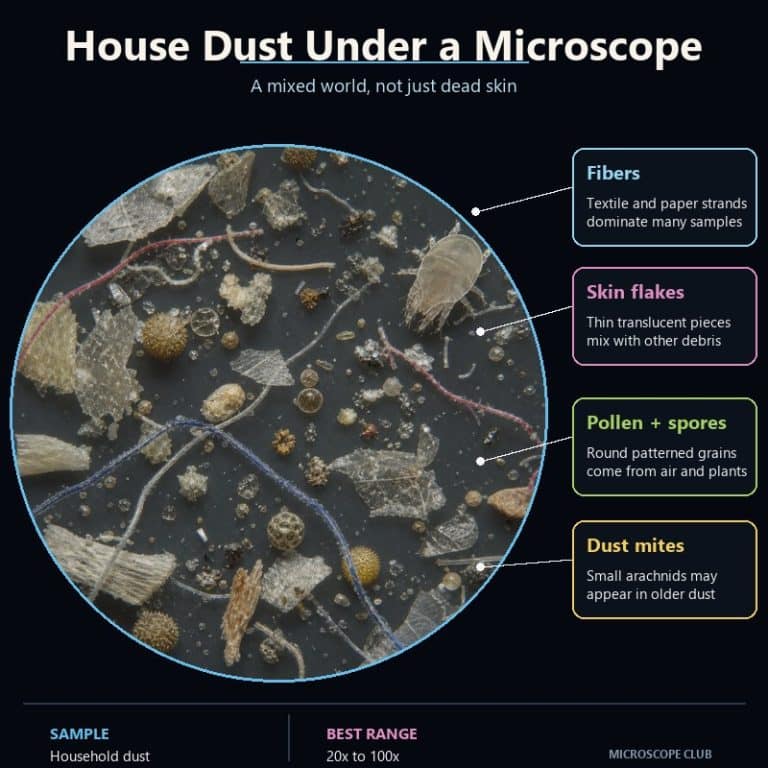
House dust under a microscope turns out to be a mixture of shed skin flakes, textile and paper fibers, pollen grains, mold spores, dust mites and their droppings, and tracked-in mineral particles. It isn’t the featureless gray fluff it looks like to the naked eye. Despite the popular myth, most of it doesn’t even come from your body. Studies show roughly 60% of household dust originates outdoors, tracked in on shoes and drifting in through windows, doors, and HVAC systems. The rest forms indoors from skin, fabric, and everyday household debris.
What House Dust Is Actually Made Of
House dust is not one thing. It is a heterogeneous mix that changes with the season, whether you have pets, how close you live to bare soil, and even which room you sample. There is no single fixed recipe — but researchers have measured its general sources closely enough to bust a few persistent myths.
A landmark modeling study by Layton and Beamer (2009), published in Environmental Science & Technology, estimated that about 60% of the dust inside an average home originates outdoors — soil tracked in on shoes, plus airborne particles that drift in through open windows, doors, and ventilation systems. The EPA’s indoor air quality research also identifies this infiltration pathway as a major route for outdoor particles entering homes. The remaining fraction forms indoors, generated by skin, textiles, cooking, and everyday wear on household materials.
| Component | What It Is | Rough Share / Source | Under the Microscope | Typical Size |
|---|---|---|---|---|
| Skin flakes | Shed dead skin cells (corneocytes) | Minor but steady, indoor source | Flat, irregular translucent plates, often stacked in sheets | 30–50 microns |
| Textile fibers | Shed cotton, wool, and synthetic threads | Usually the most abundant fibrous fraction | Twisted ribbons (cotton), scaled cylinders (wool), or smooth even rods (synthetic) | Varies; often several hundred microns long |
| Paper/cellulose fibers | Shredded paper, tissue, packaging | Minor, indoor | Short, matted, semi-translucent strands | Varies |
| Human and pet hair | Shed hair strands | Minor; higher with pets | Thick strands with a scaly cuticle and visible medulla | 50–100 microns diameter |
| Pollen | Plant reproductive grains | Seasonal, part of the outdoor fraction | Spiky or ridged spheres, often yellow | 10–100 microns |
| Mold spores | Airborne fungal reproductive cells | Minor, present year-round | Small round or oval bodies, sometimes in chains | 2–10 microns |
| Dust mites and droppings | Microscopic arthropods and their fecal pellets | Up to ~500 mites per gram of dust | Translucent globular body with eight legs; amber fecal pellets nearby | Mite: 200–300 microns; pellets: 10–40 microns |
| Mineral and soil particles | Tracked-in dirt, sand, quartz | Major share of the outdoor-origin fraction | Angular, glassy grains that flash brightly under light | Varies |
Is House Dust Really Mostly Dead Skin?
No. The claim that “80% of household dust is dead skin” is one of the most repeated facts about dust — and it isn’t supported by the research. Shed skin flakes are a real, visible component of dust, and they are the main food source for dust mites. But they are a minor fraction of the total, not the majority. The fibrous portion of most household dust is typically dominated by cotton and other textile fibers, while the fine particulate portion includes skin cells alongside mineral grains, pollen, and soot. Skin is present in every sample — it just isn’t the star of the show.
What Each Part of Dust Looks Like Under the Microscope
Under low power, a pinch of house dust looks like a gray lint ball. Bring it into focus at 100× and it explodes into a tangle of colored threads, translucent plates, and hard glassy fragments — every component with its own distinct shape.
Skin flakes show up as pale, crinkled plates — like cornflakes that have gone slightly translucent — often stacked into thin, layered sheets rather than sitting as single flat pieces. You can browse flakes of shed skin in more detail if you want a closer comparison to dandruff specifically.
Textile fibers are usually the most visually striking part of a dust sample. Cotton fibers appear as flat, twisted ribbons, kinked along their length the way a ribbon curls after you run scissors down it. Wool fibers are cylindrical, with overlapping surface scales that look like shingles on a roof. Synthetic fibers — polyester and nylon — are glassy, perfectly even rods, often dyed a shockingly bright blue or red that natural fibers rarely match. A growing share of these are now counted as microplastic fibers. All three types are strongly birefringent, meaning they light up in vivid color when viewed under polarized light. That’s one of the fastest ways to confirm a fiber is textile rather than mineral.

Paper and cellulose fibers from books, tissues, and packaging show up as shorter, matted, semi-translucent strands. They’re duller and less structured than the paper fibers you’d see under a microscope fresh off a printed page.
Human and pet hair reads as a thick strand with a visibly scaly outer cuticle and a central medulla running down the core. Pet hair often has a distinctive banded medulla pattern that separates it from human hair at a glance. Comparing human and pet hair up close side by side makes the medulla difference obvious.
Pollen grains look like yellow, studded balls — miniature sea mines with spikes, ridges, or pores across the surface, depending on the plant species. If your dust sample was taken near an open window in spring, expect to find several. A close look at pollen grains shows just how much surface detail varies species to species.

Mold spores are small, round or oval bodies, sometimes strung together in short chains, and easy to miss at low power because of their size. The CDC’s mold guidance notes that spores are airborne year-round in most climates, which is why they turn up in nearly every dust sample. For a deeper look, see mold spores and how they compare across different fungal species.
Mineral and soil particles are the hard, angular chips in the mix — sharp, glassy fragments of quartz and clay that flare bright white when you nudge the light source. That’s a sharp contrast to the soft, translucent organic material around them.
Dust Mites Under the Microscope
Dust mites are the component people are most curious — and most anxious — about. Two species dominate homes: Dermatophagoides farinae, the American house dust mite, and Dermatophagoides pteronyssinus, the European house dust mite. Both measure only 200–300 microns, right at or below the limit of naked-eye vision, so they are effectively invisible without magnification.
At 40–100×, a dust mite looks like a fat, semi-transparent grain of rice with eight stubby legs, pale and ghost-like, with its internal structures faintly visible through the translucent cuticle. Fine hairs (setae) bristle across the legs and body, and the cuticle itself has a ridged, striated texture rather than a smooth shell. Mites do not bite and are not parasites — they graze quietly on shed skin flakes, which is why they cluster wherever skin accumulates: mattresses, pillows, and upholstered furniture.
The allergy connection isn’t the mite itself — it’s what the mite leaves behind. According to the Asthma and Allergy Foundation of America, the proteins that trigger dust mite allergy (Der p 1 and Der f 1) come from the mites’ fecal pellets and shed exoskeletons. They don’t come from a bite or sting. Under the scope, those pellets appear as tiny amber-brown beads scattered around the mite’s body, each only 10–40 microns across. Populations can reach up to 500 mites per gram of dust, and a well-used mattress can harbor anywhere from 100,000 to several million.
This section covers general biology and is educational, not medical advice. If you experience ongoing allergy or asthma symptoms you suspect are related to dust mites, talk to a healthcare provider about testing and treatment options.
How to Collect and View Your Own Dust
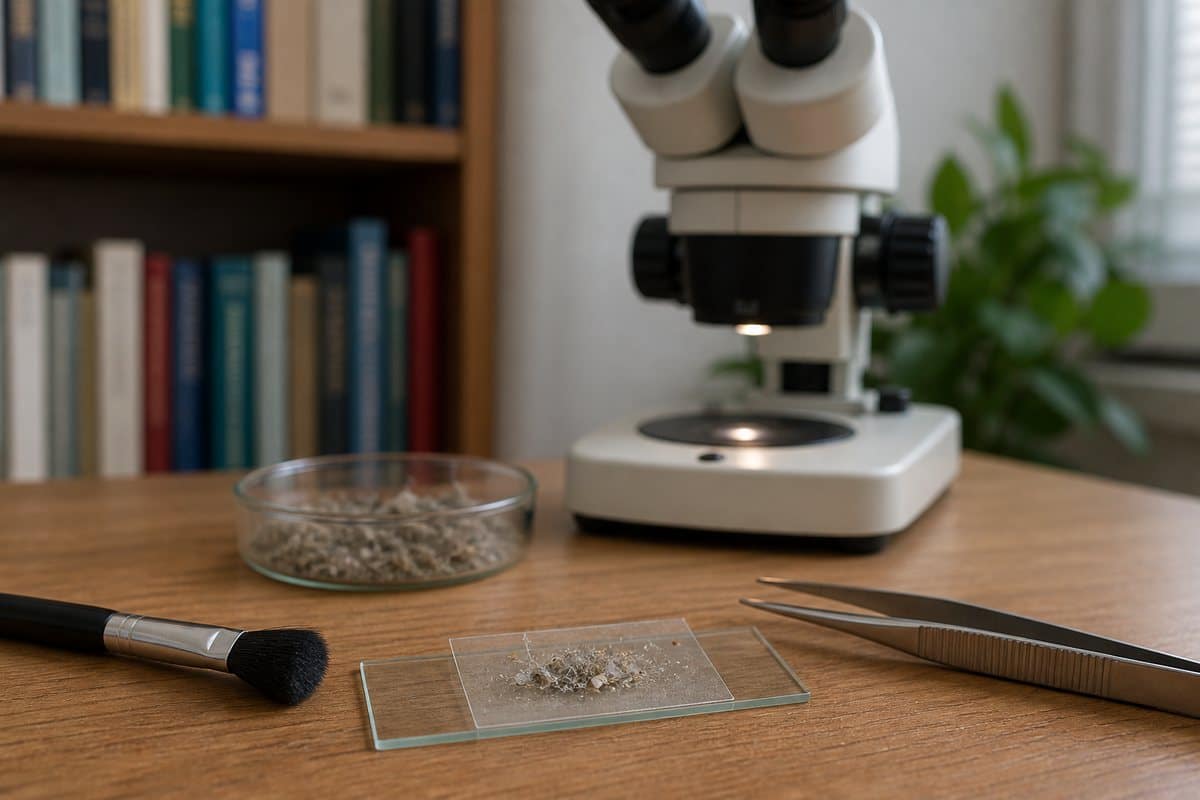
You don’t need lab equipment to sample your own house dust — just a microscope, a slide, and a source of dust that matches what you’re hoping to find.
- Choose your sample source. Windowsill or floor dust gives a broad mix of fibers, skin, pollen, and mineral grains. Mattress or pillow dust is the best source for live, moving dust mites, since that’s where they feed and breed.
- Lift the sample. Press a strip of clear tape onto the dusty surface, then stick the tape directly onto a glass slide. A tape-lift holds fine, dry particles in place far better than a wet mount. A wet mount tends to make loose dust drift and swirl away from the coverslip before you can focus.
- Spread it thin. If you’re working with a loose clump rather than a tape-lift, tease the dust apart with a probe before adding a coverslip. A thick clump just reads as a black blob under the scope — and pressing the coverslip down too hard on a live mite will crush it.
- Light it carefully. Start with low, angled light and stop down the condenser iris rather than flooding the sample with brightness. Skin flakes and mite bodies are translucent, and too much light washes out the detail that makes them recognizable. Dark-field or polarized light dramatically improves contrast on both.
- Start low, then climb. Begin at 40× to survey the whole sample and spot anything interesting, then move up to 100× or 400× to examine individual components more closely. Dust doesn’t dry out or move on its own, so there’s no rush.
Keep your sleeve and hands away from the slide while you work — stray fibers from your own clothing are a common source of contamination in a dust mount. If you’re new to slide prep in general, it helps to first prepare a slide with an easier, more forgiving specimen before attempting a tape-lift.
Best Magnification and Settings for Viewing Dust
Different dust components come into view at different magnifications, so a quick survey at low power followed by a closer look is the most efficient approach.
- 40× (survey view): whole fibers, large skin flakes, whole dust mites, and hair strands.
- 100×: mite leg structure and body shape, fiber cross-texture, pollen surface ornamentation, and skin-flake outlines.
- 400×: mold spores, fecal pellets, and fine surface detail on smaller particles.
Dark-field or polarized light is worth setting up whenever you’re viewing translucent material. It dramatically improves contrast on skin flakes and mite bodies, and it makes fibers glow with color thanks to their birefringence. That’s a fast way to separate textile fibers from mineral or synthetic look-alikes.
Frequently Asked Questions
Can you see dust mites with a normal microscope?
Yes. A basic compound or stereo microscope at 40–100× is enough to see a dust mite’s body shape, legs, and general structure. You don’t need specialized lab equipment.
What magnification do you need to see house dust clearly?
Start at 40× for a general survey, then move to 100× to identify individual components like fibers, skin flakes, and mite anatomy. Save 400× for the smallest details, such as mold spores and fecal pellets.
Do dust mites bite?
No. Dust mites don’t bite or sting. They feed on shed skin flakes, and the allergic reaction people associate with them actually comes from proteins in their fecal pellets and shed exoskeletons, not from any physical contact.
Are the tiny fibers in dust natural or synthetic?
Both. Most household dust contains a mix of natural fibers like cotton and wool alongside synthetic fibers like polyester and nylon shed from clothing, upholstery, and carpet. The synthetic share has been rising as more textiles are made from synthetic blends.
Is breathing house dust dangerous?
For most people, ordinary household dust is a minor irritant rather than a hazard. For people with dust mite or mold allergies, repeated exposure can trigger allergy or asthma symptoms. This article is educational, not medical advice — anyone with persistent respiratory symptoms should speak with a healthcare provider.
Why is there more dust in some rooms than others?
Dust accumulates fastest where there’s more fabric, foot traffic, or airflow from outside. Bedrooms with mattresses and carpet, and rooms near frequently opened doors or windows, tend to collect more than sealed, low-traffic spaces.
Conclusion
House dust under a microscope is far more interesting than its reputation suggests. Once you know what you’re looking at — skin flakes, twisted cotton ribbons, glassy synthetic fibers, spiked pollen grains, and the occasional pale, eight-legged mite — the gray fluff on a windowsill turns into a small catalog of everything that passes through your home. Some of it comes from inside your own walls, and some rides in from the world tracked through the front door.
Have you looked at your own household dust under a microscope? Tell us what you found in the comments below — especially if you spotted a live mite or something you couldn’t identify.