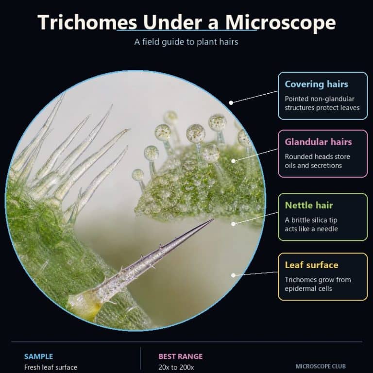
Trichomes under a microscope reveal a plant’s leaves and stems as far stranger surfaces than the naked eye suggests. Expect forests of glassy spikes, translucent balloon-headed glands, and even a tiny hypodermic needle. A trichome is simply a fine outgrowth of the plant epidermis — a plant hair — and it falls into one of two functional families: protective (non-glandular) or secretory (glandular). Grab a leaf from your kitchen or garden and a basic scope, and both types are easy to find.
The Two Types of Trichomes — Glandular vs Non-Glandular
Every trichome belongs to one of two functional families, and the split comes down to a single structural feature: whether the hair ends in a secretory head. Non-glandular trichomes are simple physical outgrowths with no head, while glandular trichomes carry one or more secretory cells on a stalk. Both types arise only from epidermal cells, which is why they’re classed separately from root hairs — a common point of confusion covered in the FAQ below.
| Feature | Non-Glandular | Glandular |
|---|---|---|
| Secretory head? | No | Yes — one or more secretory cells |
| Structure | Straight, hooked, star-shaped, branched, or shield-like | Stalk topped by a peltate (broad) or capitate (small, rounded) gland |
| Function | Reduces water loss, reflects UV, deters small insects | Secretes essential oils, resins, mucilage, or defensive compounds |
| Example | Cotton fiber (a seed trichome) | Peppermint oil gland |
Non-glandular (covering) trichomes — protection, water, UV
Non-glandular trichomes work as a physical shield rather than a chemical one. By standing off the leaf surface, they trap a thin, still layer of humid air that slows transpiration and cuts water loss. That same dense layer reflects excess sunlight and UV radiation, keeping the leaf surface cooler. It also forms a mechanical barrier that makes it harder for small insects to reach the epidermis and start feeding. Shapes vary widely — straight, hooked, star-shaped (stellate), branched (dendritic), T-shaped, or flattened into shield-like scales (peltate scales) — but none of them carry a secretory head. Cotton fiber is a non-glandular seed trichome, so if you’ve ever worn a cotton shirt, you’ve had a trichome on your back.
Glandular (secretory) trichomes — oils, resins, defense
Glandular trichomes add a working factory to the tip of the hair: a head of one or more secretory cells sitting on a stalk. That head manufactures and stores essential oils, terpenes, resins, mucilage, salts, or defensive metabolites, depending on the species. Two sub-shapes cover most of what you’ll find on common plants: peltate glands, which are short-stalked or stalkless with a broad, multicellular head built for oil storage. Capitate glands, the other sub-shape, have a distinct stalk topped by a small rounded head. The mint and cannabis examples later in this article are both variations on this same basic design.
How to See Trichomes Under a Microscope
Trichomes are three-dimensional surface structures, not thin, transparent sections, so the biggest mistake beginners make is reaching straight for a compound microscope and a wet mount. Get the setup right first and most common leaf trichomes are visible within a minute of picking the leaf.
What you need and what magnification
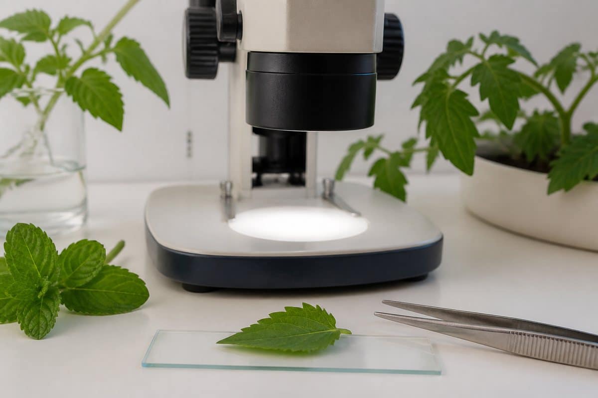
A stereo (dissecting) microscope at 10–40x is the best tool for trichomes. Its depth of field and top-down lighting let you see the hairs standing up in relief, intact and three-dimensional, which is exactly how they exist on the plant. A compound microscope at 40–400x resolves finer cellular detail — the individual cells inside a tomato gland head, for instance. But its shallow depth of field means only one thin plane stays in focus at a time. A coverslip pressed down over a wet mount can also flatten or snap the hairs before you ever get a clear view.
Preparing the leaf without crushing the hairs
Work from a fresh leaf — young leaves and stems carry the densest trichome cover, since hair density drops as tissue matures. Don’t reach for the thick middle of the leaf on a compound scope; it’s too dense and opaque for light to pass through cleanly. Instead, look at the thin leaf edge or the underside near the midrib, where trichomes sit denser and the tissue is thin enough to transmit light. If you do want a wet mount for compound viewing, avoid slamming the coverslip straight down — it flattens delicate hairs and ruptures glandular heads instantly. Use a bridged or raised coverslip (resting on two thin spacers) so the glass never touches the trichomes directly. View the leaf dry under the stereo scope first as your baseline.

Numbered viewing steps
- Pick a fresh young leaf or stem tip and handle it by the petiole — don’t rub the surface, which can smear or rupture glandular heads.
- Set the leaf under a stereo microscope first and view it dry at 10–40x with top (incident) lighting, not light shining up through the leaf.
- Scan the leaf edge and the underside along the midrib, where trichomes are densest and easiest to isolate against the background.
- If you want cellular detail on a gland head or hair tip, move to a compound microscope at 40–100x and prepare a bridged wet mount so the coverslip doesn’t crush the sample.
- Back off the condenser or switch to oblique lighting if transmitted light washes out clear, translucent hairs — most trichomes have almost no color of their own.
- Work quickly once the leaf is off the plant; a drying leaf edge causes hairs to collapse within minutes, so keep a drop of water at the leaf margin if you’re not viewing immediately.
Trichomes on Three Common Plants (Tomato, Mint, Stinging Nettle)
Trichome diversity is easiest to grasp side by side, so here’s what three plants you can find at a grocery store or garden actually show you at the eyepiece.
Tomato — the four-celled Type VI gland head
Tomato foliage carries up to seven distinct trichome types (classified as Types I through VII), a mix of glandular and non-glandular hairs, with young leaves and stems the densest. The star of the group is Type VI, a short glandular trichome topped with a four-celled globular gland head sitting on a single stalk cell. Under the scope it looks like four soap bubbles fused onto a short post. Rupture that head and you release the sticky defensive compounds responsible for the distinctive smell and tackiness of tomato leaves. A smear of that resin on your fingers after brushing a tomato plant is Type VI trichome contents, not dirt. Non-glandular Types II, III, and V fill in the surface between the glands as a forest of upright, translucent spikes.
Peppermint — peltate oil-storage glands
Peppermint and spearmint leaves carry peltate glandular trichomes. These are a disc of roughly eight secretory cells sitting beneath a raised cuticle that balloons outward as it fills with essential oil (menthol and menthone, primarily). Under the scope they look like flat, translucent blisters lying flush against the leaf surface. On an angled capitate gland, it’s more of a clear pinhead balloon on a stick. Press a coverslip onto them and they pop and deflate almost instantly, which is your cue to view mint leaves dry under a stereo scope first. Smaller capitate glandular trichomes sit alongside the peltate ones and are easy to mistake for surface debris until you get the lighting right. The smell of crushed mint leaves is literally these peltate glands emptying their oil.

Stinging nettle — the silica-tipped needle
Stinging nettle’s defining trichome is a hollow, needle-like hair with a silicified, glass-like tip sitting on a bulbous, multicellular pedestal. Under the scope it looks like a hypodermic needle balanced on a tiny onion, and the glassy tip catches the light distinctly from the surrounding tissue. On contact, that brittle tip snaps off at a bevel and injects a fluid carrying histamine, acetylcholine, serotonin, and formic acid. That fluid also carries oxalic and tartaric acids and moroidin-type peptides identified in more recent research. Nettle leaves also carry ordinary non-stinging hairs alongside the stinging ones, so not every hair you see is a hazard. Handle nettle leaves with gloves when collecting a sample — the sting is generally harmless but can irritate skin, and this is general educational information, not medical advice. Brush a nettle leaf edge-on with a gloved hand and you can watch the broken needle tips scattered across the surface under a stereo scope.
Why Cannabis Growers Check Trichomes
Cannabis trichomes that growers inspect before harvest are capitate-stalked glandular trichomes, structurally the same family as the peppermint and tomato glands described above. They manufacture cannabinoids and terpenes in the resin head instead of menthol or defensive compounds. Growers judge harvest timing by gland head color. Clear heads mean the trichome hasn’t finished producing resin, cloudy or milky heads signal peak compound concentration, and amber heads indicate the resin has begun to degrade. It’s the same basic glandular structure covered above — a stalk topped by a secretory head — just with a different secretion and a grower-specific reason to watch it closely.
What Trichomes Do for the Plant
Across both families, trichomes give a plant tools it can’t get any other way. Non-glandular hairs reduce water loss by trapping a humid boundary layer at the leaf surface and reflect excess solar and UV radiation to keep leaf temperature down. They also physically block small insects from reaching the epidermis to feed. Glandular hairs add a chemical layer of defense — secreting toxic or distasteful metabolites that deter herbivores, along with the oils and resins that give many plants their characteristic scent. Some trichomes serve dispersal instead of defense: cotton fiber, a non-glandular seed trichome, is built to catch wind and carry seeds away from the parent plant. Not every plant invests in this cover at all. Many species are glabrous, meaning smooth and hairless, because the water and UV pressures that favor trichomes don’t apply equally everywhere.
Frequently Asked Questions
Are trichomes the same as root hairs?
No. Root hairs are epidermal outgrowths too, but they form below ground on roots and are classified separately from trichomes, which occur on aerial surfaces like leaves, stems, and flowers.
Can you see trichomes without a microscope?
Often, yes — dense trichome cover shows up to the naked eye as visible fuzz or down on leaves and stems. Seeing individual hair structure or a gland head clearly, though, needs magnification.
Do all plants have trichomes?
No. Many species are glabrous, meaning naturally smooth and hairless, because they don’t face the same water-loss, UV, or herbivory pressures that favor trichome cover in other plants.
Does a hairy leaf mean a plant is unhealthy?
No — dense trichome cover, like the fuzz on tomato stems and leaves, is normal and often protective, not a sign of disease or stress.
Why are trichomes clear instead of green?
Most trichomes are translucent to clear because their cell walls carry no chlorophyll. Any color you see, such as an amber cannabis gland head, comes from the secretion inside, not the trichome structure itself.
Why do some trichomes feel brittle or glassy?
Some non-glandular trichomes, including the stinging nettle’s tip, are mineralized with silica or calcium deposits (cystoliths), which makes them rigid and prone to snapping rather than bending.
Can you see trichomes with a smartphone macro lens instead of a microscope?
A clip-on macro lens can pick up dense non-glandular hair cover and larger glandular heads like tomato’s Type VI gland. But it won’t resolve fine cellular detail — for that, a stereo or compound microscope is still necessary.
Conclusion
Trichomes turn an ordinary leaf into a study in structural diversity. Non-glandular hairs stand guard against water loss and UV, and glandular heads manufacture everything from mint oil to tomato’s sticky defense compounds. Stinging nettle’s glass-tipped needle does something closer to chemical warfare. All of it is visible with nothing more exotic than a stereo microscope and a leaf from your own kitchen or garden.
Have you looked at trichomes on a plant from your own garden or kitchen? Tell us what you found — and which plant surprised you most — in the comments below.
For more on the surface these hairs grow from, see the leaf’s epidermal layers under a microscope. If you want to compare plant hairs against an entirely different structure, human hair under a microscope is built on a completely different biology. The epidermal-peel technique used to isolate trichomes is the same one covered in onion cells under a microscope. If you’re prepping your first wet mount, how to make a wet mount slide covers the crush-caution in more depth. For another plant surface structure worth exploring next, see pollen grains under a microscope.
Sources: trichome structure and function are documented by university botany departments and peer-reviewed plant science research. This includes work on tomato Luckwill-type trichomes and nettle sting chemistry published via the National Center for Biotechnology Information. General plant epidermis and leaf anatomy background is available from Encyclopedia Britannica’s entry on trichomes. For microscopy technique guidance on depth of field and specimen mounting, see Olympus’s microscope resource center.